 Movie
Movie Controller

[English] 日本語
 Yorodumi
- PDB-3v15: Crystal structure of the Fe(II)/alpha-ketoglutarate dependent tau... -
+ Open data

- Basic information
| Entry | Database: PDB / ID: 3v15 | ||||||
|---|---|---|---|---|---|---|---|
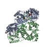
| Title | Crystal structure of the Fe(II)/alpha-ketoglutarate dependent taurine dioxygenase from Pseudomonas putida KT2440 | ||||||
 Components Components | Alpha-ketoglutarate-dependent taurine dioxygenase | ||||||
 Keywords Keywords |  OXIDOREDUCTASE / jelly roll motif / dioxygenase / alpha-ketoglutarate / Fe(II) OXIDOREDUCTASE / jelly roll motif / dioxygenase / alpha-ketoglutarate / Fe(II) | ||||||
| Function / homology |  Function and homology information Function and homology informationsulfonate dioxygenase activity / sulfur compound catabolic process / taurine dioxygenase / taurine dioxygenase activity / iron ion binding / identical protein binding / cytoplasmSimilarity search - Function | ||||||
| Biological species |  Pseudomonas putida (bacteria) Pseudomonas putida (bacteria) | ||||||
| Method | X-RAY DIFFRACTION / MOLECULAR REPLACEMENT / Resolution: 2.6 Å | ||||||
 Authors Authors | Knauer, S.H. / Dobbek, H. | ||||||
 Citation Citation | Journal: Febs J. / Year: 2012 Title: The Fe(II) / alpha-ketoglutarate dependent taurine dioxygenases from Pseudomonas putida and Escherichia coli are tetramers Authors: Knauer, S.H. / Hartl-Spiegelhauer, O. / Schwarzinger, S. / Hanzelmann, P. / Dobbek, H. | ||||||
| History |
|
- Structure visualization
| Structure viewer | Molecule: MolmilJmol/JSmol |
|---|
- Downloads & links
-Download
| PDBx/mmCIF format | 3v15.cif.gz | 392.7 KB | Display | PDBx/mmCIF format |
|---|---|---|---|---|
| PDB format | pdb3v15.ent.gz | 323.8 KB | Display | PDB format |
| PDBx/mmJSON format | 3v15.json.gz | Tree view | PDBx/mmJSON format | |
| Others |  Other downloads Other downloads |
-Validation report
| Arichive directory | https://data.pdbj.org/pub/pdb/validation_reports/v1/3v15ftp://data.pdbj.org/pub/pdb/validation_reports/v1/3v15 | HTTPS FTP |
|---|
-Related structure data
| Related structure data |  3v17C  3pvjS S: Starting model for refinement C: citing same article ( |
|---|---|
| Similar structure data |


-Links
 PDBj
PDBj- Assembly
| Deposited unit | 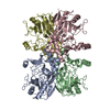
| ||||||||
|---|---|---|---|---|---|---|---|---|---|
| 1 |
| ||||||||
| Unit cell |
|
-Components
| #1: Protein | Mass: 31134.020 Da / Num. of mol.: 4 Source method: isolated from a genetically manipulated source Source: (gene. exp.) Pseudomonas putida (bacteria) / Strain: KT 2440 / Gene: PP0230, PP_0230, tauD / Plasmid: pET11a / Production host: Escherichia coli (E. coli) / Strain (production host): BL21(DE3) / References: UniProt: Q88RA3, taurine dioxygenase#2: Water | ChemComp-HOH / | Water Mass: 18.015 Da / Num. of mol.: 284 / Source method: isolated from a natural source / Formula: H2O Mass: 18.015 Da / Num. of mol.: 284 / Source method: isolated from a natural source / Formula: H2O |
|---|
-Experimental details
-Experiment
| Experiment | Method: X-RAY DIFFRACTION / Number of used crystals: 1 |
|---|
- Sample preparation
| Crystal | Density Matthews: 2.83 Å3/Da / Density % sol: 56.46 % |
|---|---|
| Crystal grow | Temperature: 289 K / Method: vapor diffusion, sitting drop / pH: 6.5 Details: reservoir solution: 20 % (w/v) PEG 5,000 MME, 0.1 M Bis Tris/HCl (pH 6.5) protein solution: 21 mg/ml protein in 25 mM Tris/HCl (pH 7.7) containing 10 mM taurine, 0.1 (NH4)2SO4 and 20 % (v/v) ...Details: reservoir solution: 20 % (w/v) PEG 5,000 MME, 0.1 M Bis Tris/HCl (pH 6.5) protein solution: 21 mg/ml protein in 25 mM Tris/HCl (pH 7.7) containing 10 mM taurine, 0.1 (NH4)2SO4 and 20 % (v/v) glycerol, VAPOR DIFFUSION, SITTING DROP, temperature 289K |
-Data collection
| Diffraction | Mean temperature: 100 K |
|---|---|
| Diffraction source | Source: ROTATING ANODE / Type: OTHER / Wavelength: 1.5418 Å |
| Detector | Type: MARRESEARCH / Detector: IMAGE PLATE / Date: Nov 20, 2006 |
| Radiation | Monochromator: osmic mirrors / Protocol: SINGLE WAVELENGTH / Monochromatic (M) / Laue (L): M / Scattering type: x-ray |
| Radiation wavelength | Wavelength: 1.5418 Å / Relative weight: 1 |
| Reflection | Resolution: 2.6→30 Å / Num. all: 44171 / Num. obs: 44029 / % possible obs: 99.7 % / Observed criterion σ(F): 2 / Observed criterion σ(I): 2 / Redundancy: 4.6 % / Rsym value: 0.088 / Net I/σ(I): 18.4 |
| Reflection shell | Resolution: 2.6→2.7 Å / Redundancy: 4.6 % / Num. unique all: 4669 / Rsym value: 0.595 / % possible all: 99.7 |
- Processing
| Software |
| ||||||||||||||||||||||||||||||||||||||||||||||||||||||||||||||||||||||||||||||||||||||||||||||||||||
|---|---|---|---|---|---|---|---|---|---|---|---|---|---|---|---|---|---|---|---|---|---|---|---|---|---|---|---|---|---|---|---|---|---|---|---|---|---|---|---|---|---|---|---|---|---|---|---|---|---|---|---|---|---|---|---|---|---|---|---|---|---|---|---|---|---|---|---|---|---|---|---|---|---|---|---|---|---|---|---|---|---|---|---|---|---|---|---|---|---|---|---|---|---|---|---|---|---|---|---|---|---|
| Refinement | Method to determine structure: MOLECULAR REPLACEMENT Starting model: PDB entry 3PVJ Resolution: 2.6→28.78 Å / SU ML: 0.38 / σ(F): 1.99 / Phase error: 23.85 / Stereochemistry target values: ML
| ||||||||||||||||||||||||||||||||||||||||||||||||||||||||||||||||||||||||||||||||||||||||||||||||||||
| Solvent computation | Shrinkage radii: 0.9 Å / VDW probe radii: 1.11 Å / Solvent model: FLAT BULK SOLVENT MODEL / Bsol: 51.583 Å2 / ksol: 0.346 e/Å3 | ||||||||||||||||||||||||||||||||||||||||||||||||||||||||||||||||||||||||||||||||||||||||||||||||||||
| Displacement parameters |
| ||||||||||||||||||||||||||||||||||||||||||||||||||||||||||||||||||||||||||||||||||||||||||||||||||||
| Refinement step | Cycle: LAST / Resolution: 2.6→28.78 Å
| ||||||||||||||||||||||||||||||||||||||||||||||||||||||||||||||||||||||||||||||||||||||||||||||||||||
| Refine LS restraints |
| ||||||||||||||||||||||||||||||||||||||||||||||||||||||||||||||||||||||||||||||||||||||||||||||||||||
| LS refinement shell |
| ||||||||||||||||||||||||||||||||||||||||||||||||||||||||||||||||||||||||||||||||||||||||||||||||||||
| Refinement TLS params. | Method: refined / Refine-ID: X-RAY DIFFRACTION
| ||||||||||||||||||||||||||||||||||||||||||||||||||||||||||||||||||||||||||||||||||||||||||||||||||||
| Refinement TLS group |
|
