 Movie
Movie Controller


+ Open data

- Basic information
| Entry | Database: PDB / ID: 1gy9 | ||||||
|---|---|---|---|---|---|---|---|
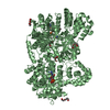
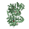
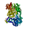
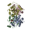
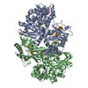
| Title | Taurine/alpha-ketoglutarate Dioxygenase from Escherichia coli | ||||||
 Components Components | ALPHA-KETOGLUTARATE-DEPENDENT TAURINE DIOXYGENASE | ||||||
 Keywords Keywords |  OXIDOREDUCTASE / TAURINE / SULPHUR METABOLISM / OXYGENASE / ALPHA-KETOGLUTARATE / TAUD / TFDA OXIDOREDUCTASE / TAURINE / SULPHUR METABOLISM / OXYGENASE / ALPHA-KETOGLUTARATE / TAUD / TFDA | ||||||
| Function / homology |  Function and homology information Function and homology informationtaurine catabolic process / taurine dioxygenase complex / taurine dioxygenase / taurine dioxygenase activity / sulfur compound metabolic process / L-ascorbic acid binding / ferrous iron binding / protein homotetramerization / identical protein binding / cytoplasmSimilarity search - Function | ||||||
| Biological species |  ESCHERICHIA COLI (E. coli) ESCHERICHIA COLI (E. coli) | ||||||
| Method | X-RAY DIFFRACTION / SYNCHROTRON / MOLECULAR REPLACEMENT / Resolution: 2.5 Å | ||||||
 Authors Authors | Elkins, J.M. / Burzlaff, N.I. / Ryle, M.J. / Lloyd, J.S. / Clifton, I.J. / Baldwin, J.E. / Hausinger, R.P. / Roach, P.L. | ||||||
 Citation Citation | Journal: Biochemistry / Year: 2002 Title: X-Ray Crystal Structure of Escherichia Coli Taurine/Alpha-Ketoglutarate Dioxygenase Complexed to Ferrous Iron and Substrates. Authors: Elkins, J.M. / Ryle, M.J. / Clifton, I.J. / Dunning Hotopp, J.C. / Lloyd, J.S. / Burzlaff, N.I. / Baldwin, J.E. / Hausinger, R.P. / Roach, P.L. | ||||||
| History |
|
- Structure visualization
| Structure viewer | Molecule: MolmilJmol/JSmol |
|---|
- Downloads & links
-Download
| PDBx/mmCIF format | 1gy9.cif.gz | 121.6 KB | Display | PDBx/mmCIF format |
|---|---|---|---|---|
| PDB format | pdb1gy9.ent.gz | 94.4 KB | Display | PDB format |
| PDBx/mmJSON format | 1gy9.json.gz | Tree view | PDBx/mmJSON format | |
| Others |  Other downloads Other downloads |
-Validation report
| Arichive directory | https://data.pdbj.org/pub/pdb/validation_reports/gy/1gy9ftp://data.pdbj.org/pub/pdb/validation_reports/gy/1gy9 | HTTPS FTP |
|---|
-Related structure data
| Related structure data |  1gqwSC S: Starting model for refinement C: citing same article ( |
|---|---|
| Similar structure data |

-Links
 PDBj
PDBj

- Assembly
| Deposited unit | 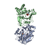
| |||||||||||||||||||||||||||
|---|---|---|---|---|---|---|---|---|---|---|---|---|---|---|---|---|---|---|---|---|---|---|---|---|---|---|---|---|
| 1 | 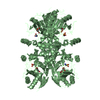
| |||||||||||||||||||||||||||
| 2 | 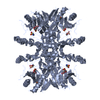
| |||||||||||||||||||||||||||
| Unit cell |
| |||||||||||||||||||||||||||
| Noncrystallographic symmetry (NCS) | NCS domain:
NCS domain segments:
NCS oper: (Code: given Matrix: (-0.85598, 0.51686, -0.01248), Vector : |
-Components
| #1: Protein | Mass: 32453.467 Da / Num. of mol.: 2 Source method: isolated from a genetically manipulated source Source: (gene. exp.) ESCHERICHIA COLI (E. coli) / Production host: ESCHERICHIA COLI (E. coli) / References: UniProt: P37610, taurine dioxygenase#2: Chemical | Iron  Mass: 55.845 Da / Num. of mol.: 3 / Source method: obtained synthetically / Formula: Fe Mass: 55.845 Da / Num. of mol.: 3 / Source method: obtained synthetically / Formula: Fe#3: Chemical | Taurine  Mass: 125.147 Da / Num. of mol.: 2 / Source method: obtained synthetically / Formula: C2H7NO3S Mass: 125.147 Da / Num. of mol.: 2 / Source method: obtained synthetically / Formula: C2H7NO3S#4: Chemical | Α-Ketoglutaric acid  Mass: 146.098 Da / Num. of mol.: 2 / Source method: obtained synthetically / Formula: C5H6O5 Mass: 146.098 Da / Num. of mol.: 2 / Source method: obtained synthetically / Formula: C5H6O5#5: Water | ChemComp-HOH / | Water Mass: 18.015 Da / Num. of mol.: 22 / Source method: isolated from a natural source / Formula: H2O Mass: 18.015 Da / Num. of mol.: 22 / Source method: isolated from a natural source / Formula: H2OCompound details | CONVERTS ALPHA KETOGLUTAR | |
|---|
-Experimental details
-Experiment
| Experiment | Method: X-RAY DIFFRACTION / Number of used crystals: 1 |
|---|
- Sample preparation
| Crystal | Density Matthews: 3 Å3/Da / Density % sol: 60 % |
|---|---|
| Crystal grow | pH: 7.5 Details: 20-28% PEG1000, 20% ETHYLENE GLYCOL, 75MM IMIDAZOLE PH7.5, PROTEIN SOLUTION LOADED WITH FE(II), ALPHA-KETOGLUTARATE, TAURINE, DITHIOTHREITOL, pH 7.50 |
-Data collection
| Diffraction | Mean temperature: 100 K |
|---|---|
| Diffraction source | Source: SYNCHROTRON / Site: ESRF  / Beamline: ID14-3 / Wavelength: 0.935 / Beamline: ID14-3 / Wavelength: 0.935 |
| Detector | Type: MARRESEARCH / Detector: CCD / Date: Dec 15, 1998 |
| Radiation | Protocol: SINGLE WAVELENGTH / Monochromatic (M) / Laue (L): M / Scattering type: x-ray |
| Radiation wavelength | Wavelength: 0.935 Å / Relative weight: 1 |
| Reflection | Resolution: 2.5→90 Å / Num. obs: 28679 / % possible obs: 99.3 % / Redundancy: 8 % / Rmerge(I) obs: 0.069 / Net I/σ(I): 7.4 |
| Reflection shell | Resolution: 2.5→2.64 Å / Redundancy: 7.8 % / Rmerge(I) obs: 0.311 / Mean I/σ(I) obs: 2.3 / % possible all: 99.1 |
- Processing
| Software |
| ||||||||||||||||||||||||||||||||||||||||||||||||||||||||||||||||||||||||||||||||||||||||||||||||||||||||||||||||||||||||||||||||||||||||||||||||||||||||||||||||||||||||||||||||||||||
|---|---|---|---|---|---|---|---|---|---|---|---|---|---|---|---|---|---|---|---|---|---|---|---|---|---|---|---|---|---|---|---|---|---|---|---|---|---|---|---|---|---|---|---|---|---|---|---|---|---|---|---|---|---|---|---|---|---|---|---|---|---|---|---|---|---|---|---|---|---|---|---|---|---|---|---|---|---|---|---|---|---|---|---|---|---|---|---|---|---|---|---|---|---|---|---|---|---|---|---|---|---|---|---|---|---|---|---|---|---|---|---|---|---|---|---|---|---|---|---|---|---|---|---|---|---|---|---|---|---|---|---|---|---|---|---|---|---|---|---|---|---|---|---|---|---|---|---|---|---|---|---|---|---|---|---|---|---|---|---|---|---|---|---|---|---|---|---|---|---|---|---|---|---|---|---|---|---|---|---|---|---|---|---|
| Refinement | Method to determine structure: MOLECULAR REPLACEMENT Starting model: PDB ENTRY 1GQW Resolution: 2.5→45.18 Å / Cor.coef. Fo:Fc: 0.918 / Cor.coef. Fo:Fc free: 0.894 / SU B: 12.51 / SU ML: 0.286 / TLS residual ADP flag: LIKELY RESIDUAL / Cross valid method: THROUGHOUT / ESU R: 0.501 / ESU R Free: 0.315 / Stereochemistry target values: MAXIMUM LIKELIHOOD Details: HYDROGENS HAVE BEEN ADDED IN THE RIDING POSITIONS RESIDUES WITH DISORDERED SIDE-CHAINS WERE MODELED AS ALANINE
| ||||||||||||||||||||||||||||||||||||||||||||||||||||||||||||||||||||||||||||||||||||||||||||||||||||||||||||||||||||||||||||||||||||||||||||||||||||||||||||||||||||||||||||||||||||||
| Solvent computation | Ion probe radii: 0.8 Å / Shrinkage radii: 0.8 Å / VDW probe radii: 1.4 Å / Solvent model: BABINET MODEL WITH MASK | ||||||||||||||||||||||||||||||||||||||||||||||||||||||||||||||||||||||||||||||||||||||||||||||||||||||||||||||||||||||||||||||||||||||||||||||||||||||||||||||||||||||||||||||||||||||
| Refinement step | Cycle: LAST / Resolution: 2.5→45.18 Å
| ||||||||||||||||||||||||||||||||||||||||||||||||||||||||||||||||||||||||||||||||||||||||||||||||||||||||||||||||||||||||||||||||||||||||||||||||||||||||||||||||||||||||||||||||||||||
| Refine LS restraints |
|