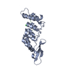
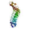
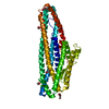
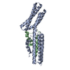
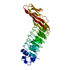
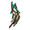
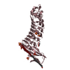
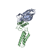
- PDB-3ui2: Crystal structure of the cpSRP54 tail bound to cpSRP43 -
+
Open data
ID or keywords:
Loading...
-
Basic information
Entry
Database: PDB / ID: 3ui2
Title
Crystal structure of the cpSRP54 tail bound to cpSRP43
Components
Signal recognition particle 43 kDa protein, chloroplastic
Signal recognition particle 54 kDa protein, chloroplastic
Keywords
TRANSPORT PROTEIN / ankyrin repeat / chromodomain / aromatic cage / signal recognition particle / protein targeting / membrane protein chaperone / chloroplast
Function / homology
Function and homology information
protein import into chloroplast thylakoid membrane / protein heterotrimerization / response to high light intensity / signal recognition particle, endoplasmic reticulum targeting / signal-recognition-particle GTPase / chloroplast envelope / 7S RNA binding / SRP-dependent cotranslational protein targeting to membrane / chloroplast stroma / chloroplast thylakoid membrane ...protein import into chloroplast thylakoid membrane / protein heterotrimerization / response to high light intensity / signal recognition particle, endoplasmic reticulum targeting / signal-recognition-particle GTPase / chloroplast envelope / 7S RNA binding / SRP-dependent cotranslational protein targeting to membrane / chloroplast stroma / chloroplast thylakoid membrane / chloroplast / disordered domain specific binding / protein-macromolecule adaptor activity / protein domain specific binding / GTPase activity / GTP binding / ATP hydrolysis activity / protein-containing complex / identical protein binding / metal ion binding / plasma membrane / cytosol Similarity search - Function
Signal recognition particle 43kDa protein / Signal recognition particle protein / Signal recognition particle, SRP54 subunit / Signal recognition particle, SRP54 subunit, M-domain / Signal recognition particle, SRP54 subunit, M-domain superfamily / Signal peptide binding domain / SRP54-type proteins GTP-binding domain signature. / Signal recognition particle SRP54, helical bundle / Signal recognition particle SRP54, N-terminal domain superfamily / SRP54-type protein, helical bundle domain ...Signal recognition particle 43kDa protein / Signal recognition particle protein / Signal recognition particle, SRP54 subunit / Signal recognition particle, SRP54 subunit, M-domain / Signal recognition particle, SRP54 subunit, M-domain superfamily / Signal peptide binding domain / SRP54-type proteins GTP-binding domain signature. / Signal recognition particle SRP54, helical bundle / Signal recognition particle SRP54, N-terminal domain superfamily / SRP54-type protein, helical bundle domain / SRP54-type protein, helical bundle domain / Signal recognition particle, SRP54 subunit, GTPase domain / SRP54-type protein, GTPase domain / SRP54-type protein, GTPase domain / Chromo domain / Chromo (CHRromatin Organisation MOdifier) domain / Chromo and chromo shadow domain profile. / OB fold (Dihydrolipoamide Acetyltransferase, E2P) - #40 / Ankyrin repeat-containing domain / Chromo/chromo shadow domain / Chromatin organization modifier domain / Chromo-like domain superfamily / Ankyrin repeat / Ankyrin repeats (3 copies) / Ankyrin repeat profile. / Ankyrin repeat region circular profile. / ankyrin repeats / Ankyrin repeat / Ankyrin repeat-containing domain superfamily / Serine Threonine Protein Phosphatase 5, Tetratricopeptide repeat / Alpha Horseshoe / OB fold (Dihydrolipoamide Acetyltransferase, E2P) / ATPases associated with a variety of cellular activities / AAA+ ATPase domain / Beta Barrel / P-loop containing nucleoside triphosphate hydrolase / Mainly Beta / Mainly Alpha Similarity search - Domain/homology
Signal recognition particle 43 kDa protein, chloroplastic / Signal recognition particle subunit SRP54, chloroplastic Similarity search - Component
In the structure databanks used in Yorodumi, some data are registered as the other names, "COVID-19 virus" and "2019-nCoV". Here are the details of the virus and the list of structure data.
Jan 31, 2019. EMDB accession codes are about to change! (news from PDBe EMDB page)
EMDB accession codes are about to change! (news from PDBe EMDB page)
The allocation of 4 digits for EMDB accession codes will soon come to an end. Whilst these codes will remain in use, new EMDB accession codes will include an additional digit and will expand incrementally as the available range of codes is exhausted. The current 4-digit format prefixed with “EMD-” (i.e. EMD-XXXX) will advance to a 5-digit format (i.e. EMD-XXXXX), and so on. It is currently estimated that the 4-digit codes will be depleted around Spring 2019, at which point the 5-digit format will come into force.
The EM Navigator/Yorodumi systems omit the EMD- prefix.
Related info.:Q: What is EMD? / ID/Accession-code notation in Yorodumi/EM Navigator
Yorodumi is a browser for structure data from EMDB, PDB, SASBDB, etc.
This page is also the successor to EM Navigator detail page, and also detail information page/front-end page for Omokage search.
The word "yorodu" (or yorozu) is an old Japanese word meaning "ten thousand". "mi" (miru) is to see.
Related info.:EMDB / PDB / SASBDB / Comparison of 3 databanks / Yorodumi Search / Aug 31, 2016. New EM Navigator & Yorodumi / Yorodumi Papers / Jmol/JSmol / Function and homology information / Changes in new EM Navigator and Yorodumi
 Movie
Movie Controller


 Open data
 Basic information
 Components
 Keywords
 TRANSPORT PROTEIN /
  Function and homology information
 Authors
 Citation
 Structure visualization
 Downloads & links
 Other downloads
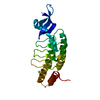
 PDBj
PDBj

 Assembly
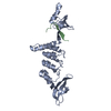

 Sample preparation
 / Beamline: ID14-2 / Wavelength: 0.933 Å
 Processing