 Movie
Movie Controller

[English] 日本語
 Yorodumi
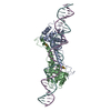
- PDB-3uec: Crystal structure of human Survivin bound to histone H3 phosphory... -
+ Open data

- Basic information
| Entry | Database: PDB / ID: 3uec | ||||||
|---|---|---|---|---|---|---|---|
| Title | Crystal structure of human Survivin bound to histone H3 phosphorylated on threonine-3. | ||||||
 Components Components |
| ||||||
 Keywords Keywords |  CELL CYCLE / zinc finger / phosphorylated threonine / BIR DOMAIN / CHROMOSOMAL PASSENGER COMPLEX / CELL DIVISION / MITOSIS CELL CYCLE / zinc finger / phosphorylated threonine / BIR DOMAIN / CHROMOSOMAL PASSENGER COMPLEX / CELL DIVISION / MITOSIS | ||||||
| Function / homology |  Function and homology informationsurvivin complex / establishment of chromosome localization / positive regulation of mitotic sister chromatid separation / positive regulation of exit from mitosis / positive regulation of mitotic cytokinesis / positive regulation of mitotic cell cycle spindle assembly checkpoint / mitotic spindle midzone assembly / positive regulation of attachment of mitotic spindle microtubules to kinetochore / interphase microtubule organizing center / protein-containing complex localization ...survivin complex / establishment of chromosome localization / positive regulation of mitotic sister chromatid separation / positive regulation of exit from mitosis / positive regulation of mitotic cytokinesis / positive regulation of mitotic cell cycle spindle assembly checkpoint / mitotic spindle midzone assembly / positive regulation of attachment of mitotic spindle microtubules to kinetochore / interphase microtubule organizing center / protein-containing complex localization / chromosome passenger complex / cobalt ion binding / cysteine-type endopeptidase inhibitor activity / nuclear chromosome / mitotic spindle assembly checkpoint signaling / TP53 regulates transcription of several additional cell death genes whose specific roles in p53-dependent apoptosis remain uncertain / cysteine-type endopeptidase inhibitor activity involved in apoptotic process / SUMOylation of DNA replication proteins / mitotic cytokinesis / chromosome, centromeric region / mitotic spindle assembly / Amplification of signal from unattached kinetochores via a MAD2 inhibitory signal / cytoplasmic microtubule / Mitotic Prometaphase / EML4 and NUDC in mitotic spindle formation / Resolution of Sister Chromatid Cohesion / centriole / positive regulation of mitotic cell cycle / tubulin binding / RHO GTPases Activate Formins / spindle microtubule / sensory perception of sound / negative regulation of cysteine-type endopeptidase activity involved in apoptotic process / kinetochore / spindle / small GTPase binding / Separation of Sister Chromatids / microtubule cytoskeleton / G2/M transition of mitotic cell cycle / mitotic cell cycle / Neddylation / midbody / protein-folding chaperone binding / microtubule binding / Interleukin-4 and Interleukin-13 signaling / microtubule / protein heterodimerization activity / cell division / protein phosphorylation / negative regulation of DNA-templated transcription / apoptotic process / positive regulation of cell population proliferation / negative regulation of apoptotic process / enzyme binding / protein homodimerization activity / protein-containing complex / zinc ion binding / nucleoplasm / identical protein binding / metal ion binding / nucleus / cytosol / cytoplasm Function and homology informationsurvivin complex / establishment of chromosome localization / positive regulation of mitotic sister chromatid separation / positive regulation of exit from mitosis / positive regulation of mitotic cytokinesis / positive regulation of mitotic cell cycle spindle assembly checkpoint / mitotic spindle midzone assembly / positive regulation of attachment of mitotic spindle microtubules to kinetochore / interphase microtubule organizing center / protein-containing complex localization ...survivin complex / establishment of chromosome localization / positive regulation of mitotic sister chromatid separation / positive regulation of exit from mitosis / positive regulation of mitotic cytokinesis / positive regulation of mitotic cell cycle spindle assembly checkpoint / mitotic spindle midzone assembly / positive regulation of attachment of mitotic spindle microtubules to kinetochore / interphase microtubule organizing center / protein-containing complex localization / chromosome passenger complex / cobalt ion binding / cysteine-type endopeptidase inhibitor activity / nuclear chromosome / mitotic spindle assembly checkpoint signaling / TP53 regulates transcription of several additional cell death genes whose specific roles in p53-dependent apoptosis remain uncertain / cysteine-type endopeptidase inhibitor activity involved in apoptotic process / SUMOylation of DNA replication proteins / mitotic cytokinesis / chromosome, centromeric region / mitotic spindle assembly / Amplification of signal from unattached kinetochores via a MAD2 inhibitory signal / cytoplasmic microtubule / Mitotic Prometaphase / EML4 and NUDC in mitotic spindle formation / Resolution of Sister Chromatid Cohesion / centriole / positive regulation of mitotic cell cycle / tubulin binding / RHO GTPases Activate Formins / spindle microtubule / sensory perception of sound / negative regulation of cysteine-type endopeptidase activity involved in apoptotic process / kinetochore / spindle / small GTPase binding / Separation of Sister Chromatids / microtubule cytoskeleton / G2/M transition of mitotic cell cycle / mitotic cell cycle / Neddylation / midbody / protein-folding chaperone binding / microtubule binding / Interleukin-4 and Interleukin-13 signaling / microtubule / protein heterodimerization activity / cell division / protein phosphorylation / negative regulation of DNA-templated transcription / apoptotic process / positive regulation of cell population proliferation / negative regulation of apoptotic process / enzyme binding / protein homodimerization activity / protein-containing complex / zinc ion binding / nucleoplasm / identical protein binding / metal ion binding / nucleus / cytosol / cytoplasmSimilarity search - Function | ||||||
| Biological species |  Homo sapiens (human) Homo sapiens (human) | ||||||
| Method | X-RAY DIFFRACTION / SYNCHROTRON / SAD / Resolution: 2.18 Å | ||||||
 Authors Authors | Niedzialkowska, E. / Porebski, P.J. / Cooper, D.R. / Chruszcz, M. / Wang, F. / Higgins, J.M. / Stukenberg, P.T. / Minor, W. | ||||||
 Citation Citation | Journal: Mol.Biol.Cell / Year: 2012 Title: Molecular basis for phosphospecific recognition of histone H3 tails by Survivin paralogues at inner centromeres. Authors: Niedzialkowska, E. / Wang, F. / Porebski, P.J. / Minor, W. / Higgins, J.M. / Stukenberg, P.T. | ||||||
| History |
| ||||||
| Remark 650 | HELIX DETERMINATION METHOD: AUTHOR DETERMINED | ||||||
| Remark 700 | SHEET DETERMINATION METHOD: AUTHOR DETERMINED |
- Structure visualization
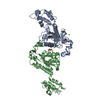
| Structure viewer | Molecule: MolmilJmol/JSmol |
|---|
- Downloads & links
-Download
| PDBx/mmCIF format | 3uec.cif.gz | 71.4 KB | Display | PDBx/mmCIF format |
|---|---|---|---|---|
| PDB format | pdb3uec.ent.gz | 57 KB | Display | PDB format |
| PDBx/mmJSON format | 3uec.json.gz | Tree view | PDBx/mmJSON format | |
| Others |  Other downloads Other downloads |
-Validation report
| Arichive directory | https://data.pdbj.org/pub/pdb/validation_reports/ue/3uecftp://data.pdbj.org/pub/pdb/validation_reports/ue/3uec | HTTPS FTP |
|---|
-Related structure data
| Related structure data | 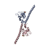 3uedC 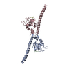 3ueeC  3uefC  3uegC 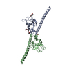 3uehC  3ueiC C: citing same article ( |
|---|---|
| Similar structure data |

-Links
 PDBj
PDBj


- Assembly
| Deposited unit | 
| ||||||||
|---|---|---|---|---|---|---|---|---|---|
| 1 | 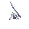
| ||||||||
| Unit cell |
| ||||||||
| Components on special symmetry positions |
|
-Components
-Protein / Protein/peptide , 2 types, 2 molecules AB
| #1: Protein | Mass: 16826.127 Da / Num. of mol.: 1 / Mutation: E129K Source method: isolated from a genetically manipulated source Source: (gene. exp.) Homo sapiens (human) / Gene: API4, BIRC5, IAP4 / Plasmid: p8HIS / Production host:  Escherichia coli (E. coli) / Strain (production host): BL21 CodonPlus(DE3)-RIL / References: UniProt: O15392 Escherichia coli (E. coli) / Strain (production host): BL21 CodonPlus(DE3)-RIL / References: UniProt: O15392 |
|---|---|
| #2: Protein/peptide | Mass: 556.550 Da / Num. of mol.: 1 / Source method: obtained synthetically |
-Non-polymers , 7 types, 60 molecules 


| #3: Chemical | ChemComp-ZN /  Mass: 65.409 Da / Num. of mol.: 1 / Source method: obtained synthetically / Formula: Zn Mass: 65.409 Da / Num. of mol.: 1 / Source method: obtained synthetically / Formula: Zn | ||||||||||
|---|---|---|---|---|---|---|---|---|---|---|---|
| #4: Chemical | Ethylene glycol Mass: 62.068 Da / Num. of mol.: 2 / Source method: obtained synthetically / Formula: C2H6O2 Mass: 62.068 Da / Num. of mol.: 2 / Source method: obtained synthetically / Formula: C2H6O2#5: Chemical | ChemComp-PG4 / | Polyethylene glycol Mass: 194.226 Da / Num. of mol.: 1 / Source method: obtained synthetically / Formula: C8H18O5 / Comment: precipitant*YM Mass: 194.226 Da / Num. of mol.: 1 / Source method: obtained synthetically / Formula: C8H18O5 / Comment: precipitant*YM#6: Chemical | ChemComp-1PE / | Polyethylene glycol Mass: 238.278 Da / Num. of mol.: 1 / Source method: obtained synthetically / Formula: C10H22O6 / Comment: precipitant*YM Mass: 238.278 Da / Num. of mol.: 1 / Source method: obtained synthetically / Formula: C10H22O6 / Comment: precipitant*YM#7: Chemical | ChemComp-ACT / | Acetate Mass: 59.044 Da / Num. of mol.: 1 / Source method: obtained synthetically / Formula: C2H3O2 Mass: 59.044 Da / Num. of mol.: 1 / Source method: obtained synthetically / Formula: C2H3O2#8: Chemical | ChemComp-PEG / | Diethylene glycol Mass: 106.120 Da / Num. of mol.: 1 / Source method: obtained synthetically / Formula: C4H10O3 Mass: 106.120 Da / Num. of mol.: 1 / Source method: obtained synthetically / Formula: C4H10O3#9: Water | ChemComp-HOH / | WaterMass: 18.015 Da / Num. of mol.: 53 / Source method: isolated from a natural source / Formula: H2O |
-Details
| Sequence details | E129K REPRESENTS |
|---|
-Experimental details
-Experiment
| Experiment | Method: X-RAY DIFFRACTION / Number of used crystals: 1 |
|---|
- Sample preparation
| Crystal | Density Matthews: 3.11 Å3/Da / Density % sol: 60.41 % |
|---|---|
| Crystal grow | Temperature: 289 K / Method: vapor diffusion, hanging drop / pH: 7.5 Details: 12% peg 8000; O.1 M MOPS 7.5; 0.1 M Magnesium acetate tetrahydrate , VAPOR DIFFUSION, HANGING DROP, temperature 289K |
-Data collection
| Diffraction | Mean temperature: 100 K |
|---|---|
| Diffraction source | Source: SYNCHROTRON / Site: APS  / Beamline: 19-ID / Wavelength: 0.9792 Å / Beamline: 19-ID / Wavelength: 0.9792 Å |
| Detector | Type: ADSC QUANTUM 315r / Detector: CCD / Date: Nov 12, 2010 / Details: mirrors |
| Radiation | Monochromator: Si 111 channel / Protocol: SINGLE WAVELENGTH / Monochromatic (M) / Laue (L): M / Scattering type: x-ray |
| Radiation wavelength | Wavelength: 0.9792 Å / Relative weight: 1 |
| Reflection | Resolution: 2.18→50 Å / Num. all: 11616 / Num. obs: 11616 / % possible obs: 99.9 % / Observed criterion σ(F): 0 / Observed criterion σ(I): -3 / Redundancy: 3.8 % / Biso Wilson estimate: 44.3 Å2 / Rmerge(I) obs: 0.067 / Net I/σ(I): 24.2 |
| Reflection shell | Resolution: 2.18→2.22 Å / Redundancy: 7.2 % / Rmerge(I) obs: 0.776 / Mean I/σ(I) obs: 2.3 / Num. unique all: 586 / % possible all: 100 |
- Processing
| Software |
| ||||||||||||||||||||||||||||||||||||||||||||||||||||||||||||||||||||||||||||||||||||||||||||||||||||
|---|---|---|---|---|---|---|---|---|---|---|---|---|---|---|---|---|---|---|---|---|---|---|---|---|---|---|---|---|---|---|---|---|---|---|---|---|---|---|---|---|---|---|---|---|---|---|---|---|---|---|---|---|---|---|---|---|---|---|---|---|---|---|---|---|---|---|---|---|---|---|---|---|---|---|---|---|---|---|---|---|---|---|---|---|---|---|---|---|---|---|---|---|---|---|---|---|---|---|---|---|---|
| Refinement | Method to determine structure: SAD / Resolution: 2.18→50 Å / Cor.coef. Fo:Fc: 0.956 / Cor.coef. Fo:Fc free: 0.957 / SU B: 9.5 / SU ML: 0.13 / Cross valid method: THROUGHOUT / σ(F): 0 / σ(I): 0 / ESU R Free: 0.159 Stereochemistry target values: MAXIMUM LIKELIHOOD WITH PHASES Details: HYDROGENS HAVE BEEN ADDED IN THE RIDING POSITIONS
| ||||||||||||||||||||||||||||||||||||||||||||||||||||||||||||||||||||||||||||||||||||||||||||||||||||
| Solvent computation | Ion probe radii: 0.8 Å / Shrinkage radii: 0.8 Å / VDW probe radii: 1.2 Å / Solvent model: MASK | ||||||||||||||||||||||||||||||||||||||||||||||||||||||||||||||||||||||||||||||||||||||||||||||||||||
| Displacement parameters | Biso mean: 56.691 Å2
| ||||||||||||||||||||||||||||||||||||||||||||||||||||||||||||||||||||||||||||||||||||||||||||||||||||
| Refinement step | Cycle: LAST / Resolution: 2.18→50 Å
| ||||||||||||||||||||||||||||||||||||||||||||||||||||||||||||||||||||||||||||||||||||||||||||||||||||
| Refine LS restraints |
| ||||||||||||||||||||||||||||||||||||||||||||||||||||||||||||||||||||||||||||||||||||||||||||||||||||
| LS refinement shell | Resolution: 2.18→2.237 Å / Total num. of bins used: 20
| ||||||||||||||||||||||||||||||||||||||||||||||||||||||||||||||||||||||||||||||||||||||||||||||||||||
| Refinement TLS params. | Method: refined / Refine-ID: X-RAY DIFFRACTION
| ||||||||||||||||||||||||||||||||||||||||||||||||||||||||||||||||||||||||||||||||||||||||||||||||||||
| Refinement TLS group |
|
