 Movie
Movie Controller

[English] 日本語
 Yorodumi
- PDB-6y42: Crystal Structure of RsrR complexed to a 39 basepair DNA fragment... -
+ Open data

- Basic information
| Entry | Database: PDB / ID: 6y42 | |||||||||
|---|---|---|---|---|---|---|---|---|---|---|
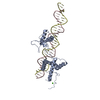
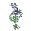
| Title | Crystal Structure of RsrR complexed to a 39 basepair DNA fragment of the rsrR promoter | |||||||||
 Components Components |
| |||||||||
 Keywords Keywords |  TRANSCRIPTION / REDOX SENSOR / IRON SULFUR CLUSTER / DNA COMPLEX TRANSCRIPTION / REDOX SENSOR / IRON SULFUR CLUSTER / DNA COMPLEX | |||||||||
| Function / homology |  Function and homology information Function and homology informationTranscription regulator Rrf2-type, conserved site / Rrf2-type HTH domain signature. / Transcription regulator Rrf2 / Iron-dependent Transcriptional regulator / Rrf2-type HTH domain profile. / Winged helix DNA-binding domain superfamily / Winged helix-like DNA-binding domain superfamily Similarity search - Domain/homology | |||||||||
| Biological species |  Streptomyces venezuelae (bacteria)Streptomyces venezuelae ATCC 10712 (bacteria) Streptomyces venezuelae (bacteria)Streptomyces venezuelae ATCC 10712 (bacteria) | |||||||||
| Method | X-RAY DIFFRACTION / SYNCHROTRON / MOLECULAR REPLACEMENT / Resolution: 4.3 Å | |||||||||
 Authors Authors | Volbeda, A. / Fontecilla-Camps, J.C. | |||||||||
| Funding support |  France, 2items France, 2items
| |||||||||
 Citation Citation | Journal: J.Am.Chem.Soc. / Year: 2020 Title: Electron and Proton Transfers Modulate DNA Binding by the Transcription Regulator RsrR. Authors: Crack, J.C. / Amara, P. / Volbeda, A. / Mouesca, J.M. / Rohac, R. / Pellicer Martinez, M.T. / Huang, C.Y. / Gigarel, O. / Rinaldi, C. / Le Brun, N.E. / Fontecilla-Camps, J.C. #1: Journal: J. Am. Chem. Soc. / Year: 2019Title: Crystal Structure of the Transcription Regulator RsrR Reveals a [2Fe-2S] Cluster Coordinated by Cys, Glu, and His Residues. Authors: Volbeda, A. / Martinez, M.T.P. / Crack, J.C. / Amara, P. / Gigarel, O. / Munnoch, J.T. / Hutchings, M.I. / Darnault, C. / Le Brun, N.E. / Fontecilla-Camps, J.C. | |||||||||
| History |
|
- Structure visualization
| Structure viewer | Molecule: MolmilJmol/JSmol |
|---|
- Downloads & links
-Download
| PDBx/mmCIF format | 6y42.cif.gz | 113.7 KB | Display | PDBx/mmCIF format |
|---|---|---|---|---|
| PDB format | pdb6y42.ent.gz | 82.4 KB | Display | PDB format |
| PDBx/mmJSON format | 6y42.json.gz | Tree view | PDBx/mmJSON format | |
| Others |  Other downloads Other downloads |
-Validation report
| Arichive directory | https://data.pdbj.org/pub/pdb/validation_reports/y4/6y42ftp://data.pdbj.org/pub/pdb/validation_reports/y4/6y42 | HTTPS FTP |
|---|
-Related structure data
| Related structure data | 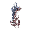 6y45C 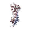 6hsdS S: Starting model for refinement C: citing same article ( |
|---|---|
| Similar structure data |


-Links
 PDBj
PDBj


- Assembly
| Deposited unit | 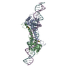
| ||||||||
|---|---|---|---|---|---|---|---|---|---|
| 1 |
| ||||||||
| Unit cell |
|
-Components
| #1: Protein | Mass: 17385.975 Da / Num. of mol.: 2 Source method: isolated from a genetically manipulated source Source: (gene. exp.) Streptomyces venezuelae (strain ATCC 10712 / CBS 650.69 / DSM 40230 / JCM 4526 / NBRC 13096 / PD 04745) (bacteria)Gene: SVEN_6563 / Plasmid: pGS-21a / Cell line (production host): BL21(DE3) Production host: Escherichia coli 'BL21-Gold(DE3)pLysS AG' (bacteria)References: UniProt: F2RGC9 #2: DNA chain | | Mass: 12127.805 Da / Num. of mol.: 1 / Source method: obtained synthetically Source: (synth.) Streptomyces venezuelae ATCC 10712 (bacteria)#3: DNA chain | | Mass: 11878.648 Da / Num. of mol.: 1 / Source method: obtained synthetically Source: (synth.) Streptomyces venezuelae ATCC 10712 (bacteria)#4: Chemical | Iron–sulfur cluster  Mass: 175.820 Da / Num. of mol.: 2 / Source method: obtained synthetically / Formula: Fe2S2 / Feature type: SUBJECT OF INVESTIGATION Mass: 175.820 Da / Num. of mol.: 2 / Source method: obtained synthetically / Formula: Fe2S2 / Feature type: SUBJECT OF INVESTIGATIONHas ligand of interest | Y | |
|---|
-Experimental details
-Experiment
| Experiment | Method: X-RAY DIFFRACTION / Number of used crystals: 1 |
|---|
- Sample preparation
| Crystal | Density Matthews: 3.5 Å3/Da / Density % sol: 64.86 % |
|---|---|
| Crystal grow | Temperature: 298 K / Method: vapor diffusion Details: NaCl, MgCl2, CaCl2, PEG1000, PEG3350, MES, anaerobic (in glove box) |
-Data collection
| Diffraction | Mean temperature: 100 K / Serial crystal experiment: N |
|---|---|
| Diffraction source | Source: SYNCHROTRON / Site: SLS  / Beamline: X06SA / Wavelength: 1.00003 Å / Beamline: X06SA / Wavelength: 1.00003 Å |
| Detector | Type: DECTRIS EIGER X 16M / Detector: PIXEL / Date: May 31, 2019 |
| Radiation | Protocol: SINGLE WAVELENGTH / Monochromatic (M) / Laue (L): M / Scattering type: x-ray |
| Radiation wavelength | Wavelength: 1.00003 Å / Relative weight: 1 |
| Reflection | Resolution: 4.3→49.34 Å / Num. obs: 6038 / % possible obs: 99.5 % / Redundancy: 5.8 % / CC1/2: 0.995 / Rmerge(I) obs: 0.087 / Rpim(I) all: 0.057 / Rrim(I) all: 0.104 / Net I/σ(I): 8.8 |
| Reflection shell | Resolution: 4.3→4.45 Å / Rmerge(I) obs: 2.13 / Mean I/σ(I) obs: 0.9 / Num. unique obs: 3563 / CC1/2: 0.36 / Rpim(I) all: 0.988 / Rrim(I) all: 2.53 |
- Processing
| Software |
| ||||||||||||||||||||||||||||||||||||||||||||||||||||||||||||
|---|---|---|---|---|---|---|---|---|---|---|---|---|---|---|---|---|---|---|---|---|---|---|---|---|---|---|---|---|---|---|---|---|---|---|---|---|---|---|---|---|---|---|---|---|---|---|---|---|---|---|---|---|---|---|---|---|---|---|---|---|---|
| Refinement | Method to determine structure: MOLECULAR REPLACEMENT Starting model: 6hsd Resolution: 4.3→49.34 Å / Cor.coef. Fo:Fc: 0.93 / Cor.coef. Fo:Fc free: 0.939 / SU B: 132.744 / SU ML: 1.358 / Cross valid method: THROUGHOUT / σ(F): 0 / ESU R Free: 1.077 Details: HYDROGENS HAVE BEEN ADDED IN THE RIDING POSITIONS U VALUES : REFINED INDIVIDUALLY
| ||||||||||||||||||||||||||||||||||||||||||||||||||||||||||||
| Solvent computation | Ion probe radii: 0.8 Å / Shrinkage radii: 0.8 Å / VDW probe radii: 1.2 Å | ||||||||||||||||||||||||||||||||||||||||||||||||||||||||||||
| Displacement parameters | Biso max: 387.16 Å2 / Biso mean: 253.122 Å2 / Biso min: 185.85 Å2
| ||||||||||||||||||||||||||||||||||||||||||||||||||||||||||||
| Refinement step | Cycle: final / Resolution: 4.3→49.34 Å
| ||||||||||||||||||||||||||||||||||||||||||||||||||||||||||||
| Refine LS restraints |
| ||||||||||||||||||||||||||||||||||||||||||||||||||||||||||||
| LS refinement shell | Resolution: 4.3→4.411 Å / Rfactor Rfree error: 0 / Total num. of bins used: 20
|
