 Movie
Movie Controller

[English] 日本語
 Yorodumi
- PDB-3u7f: Crystal structure of mPNKP catalytic fragment (D170A) bound to si... -
+ Open data

- Basic information
| Entry | Database: PDB / ID: 3u7f | ||||||
|---|---|---|---|---|---|---|---|
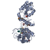
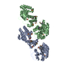
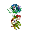
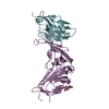
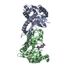
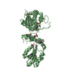
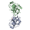
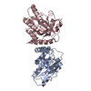
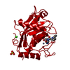
| Title | Crystal structure of mPNKP catalytic fragment (D170A) bound to single-stranded DNA (TCCTCp) | ||||||
 Components Components |
| ||||||
 Keywords Keywords |  HYDROLASE / TRANSFERASE/DNA / protein-DNA complex / HAD family / pnkp / DNA repair / phosphatase / TRANSFERASE-DNA complex HYDROLASE / TRANSFERASE/DNA / protein-DNA complex / HAD family / pnkp / DNA repair / phosphatase / TRANSFERASE-DNA complex | ||||||
| Function / homology |  Function and homology informationpolynucleotide 3'-phosphatase / polynucleotide 5'-hydroxyl-kinase / polynucleotide 3'-phosphatase activity / ATP-dependent polydeoxyribonucleotide 5'-hydroxyl-kinase activity / APEX1-Independent Resolution of AP Sites via the Single Nucleotide Replacement Pathway / DNA ligation involved in DNA repair / SCF ubiquitin ligase complex / positive regulation of double-strand break repair via nonhomologous end joining / protein K63-linked ubiquitination / positive regulation of telomere capping ...polynucleotide 3'-phosphatase / polynucleotide 5'-hydroxyl-kinase / polynucleotide 3'-phosphatase activity / ATP-dependent polydeoxyribonucleotide 5'-hydroxyl-kinase activity / APEX1-Independent Resolution of AP Sites via the Single Nucleotide Replacement Pathway / DNA ligation involved in DNA repair / SCF ubiquitin ligase complex / positive regulation of double-strand break repair via nonhomologous end joining / protein K63-linked ubiquitination / positive regulation of telomere capping / ubiquitin-like ligase-substrate adaptor activity / positive regulation of telomere maintenance via telomerase / double-strand break repair via nonhomologous end joining / site of double-strand break / double-stranded DNA binding / response to oxidative stress / phosphorylation / DNA repair / DNA damage response / nucleolus / nucleoplasm / ATP binding / nucleus Function and homology informationpolynucleotide 3'-phosphatase / polynucleotide 5'-hydroxyl-kinase / polynucleotide 3'-phosphatase activity / ATP-dependent polydeoxyribonucleotide 5'-hydroxyl-kinase activity / APEX1-Independent Resolution of AP Sites via the Single Nucleotide Replacement Pathway / DNA ligation involved in DNA repair / SCF ubiquitin ligase complex / positive regulation of double-strand break repair via nonhomologous end joining / protein K63-linked ubiquitination / positive regulation of telomere capping ...polynucleotide 3'-phosphatase / polynucleotide 5'-hydroxyl-kinase / polynucleotide 3'-phosphatase activity / ATP-dependent polydeoxyribonucleotide 5'-hydroxyl-kinase activity / APEX1-Independent Resolution of AP Sites via the Single Nucleotide Replacement Pathway / DNA ligation involved in DNA repair / SCF ubiquitin ligase complex / positive regulation of double-strand break repair via nonhomologous end joining / protein K63-linked ubiquitination / positive regulation of telomere capping / ubiquitin-like ligase-substrate adaptor activity / positive regulation of telomere maintenance via telomerase / double-strand break repair via nonhomologous end joining / site of double-strand break / double-stranded DNA binding / response to oxidative stress / phosphorylation / DNA repair / DNA damage response / nucleolus / nucleoplasm / ATP binding / nucleusSimilarity search - Function | ||||||
| Biological species |  Mus musculus (house mouse) Mus musculus (house mouse) | ||||||
| Method | X-RAY DIFFRACTION / SYNCHROTRON / Resolution: 1.8 Å | ||||||
 Authors Authors | Coquelle, N. / Havali, Z. / Bernstein, N. / Green, R. / Glover, J.N.M. | ||||||
 Citation Citation | Journal: Proc.Natl.Acad.Sci.USA / Year: 2011 Title: Structural basis for the phosphatase activity of polynucleotide kinase/phosphatase on single- and double-stranded DNA substrates. Authors: Coquelle, N. / Havali-Shahriari, Z. / Bernstein, N. / Green, R. / Glover, J.N. #1: Journal: Mol.Cell / Year: 2005Title: The molecular architecture of the mammalian DNA repair enzyme, polynucleotide kinase. Authors: Bernstein, N.K. / Williams, R.S. / Rakovszky, M.L. / Cui, D. / Green, R. / Karimi-Busheri, F. / Mani, R.S. / Galicia, S. / Koch, C.A. / Cass, C.E. / Durocher, D. / Weinfeld, M. / Glover, J.N. | ||||||
| History |
|
- Structure visualization
| Structure viewer | Molecule: MolmilJmol/JSmol |
|---|
- Downloads & links
-Download
| PDBx/mmCIF format | 3u7f.cif.gz | 223.6 KB | Display | PDBx/mmCIF format |
|---|---|---|---|---|
| PDB format | pdb3u7f.ent.gz | 188.5 KB | Display | PDB format |
| PDBx/mmJSON format | 3u7f.json.gz | Tree view | PDBx/mmJSON format | |
| Others |  Other downloads Other downloads |
-Validation report
| Arichive directory | https://data.pdbj.org/pub/pdb/validation_reports/u7/3u7fftp://data.pdbj.org/pub/pdb/validation_reports/u7/3u7f | HTTPS FTP |
|---|
-Related structure data


-Links
 PDBj
PDBj

- Assembly
| Deposited unit | 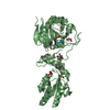
| ||||||||
|---|---|---|---|---|---|---|---|---|---|
| 1 |
| ||||||||
| Unit cell |
|
-Components
-Protein / DNA chain , 2 types, 2 molecules BX
| #1: Protein | Mass: 42378.699 Da / Num. of mol.: 1 / Mutation: D170A Source method: isolated from a genetically manipulated source Source: (gene. exp.) Mus musculus (house mouse) / Gene: Pnkp / Plasmid: pet19 / Production host:  Escherichia coli (E. coli) / Strain (production host): BL21(DE3) Escherichia coli (E. coli) / Strain (production host): BL21(DE3)References: UniProt: Q9JLV6, polynucleotide 3'-phosphatase, polynucleotide 5'-hydroxyl-kinase |
|---|---|
| #2: DNA chain | Mass: 1430.974 Da / Num. of mol.: 1 / Source method: obtained synthetically / Details: DNA substrate of PNKP phosphatase domain |
-Non-polymers , 4 types, 416 molecules 


| #3: Chemical | ChemComp-MG /  Mass: 24.305 Da / Num. of mol.: 1 / Source method: obtained synthetically / Formula: Mg Mass: 24.305 Da / Num. of mol.: 1 / Source method: obtained synthetically / Formula: Mg | ||||
|---|---|---|---|---|---|
| #4: Chemical | ChemComp-GOL / Glycerol Mass: 92.094 Da / Num. of mol.: 4 / Source method: obtained synthetically / Formula: C3H8O3 Mass: 92.094 Da / Num. of mol.: 4 / Source method: obtained synthetically / Formula: C3H8O3#5: Chemical | Phosphate Mass: 94.971 Da / Num. of mol.: 2 / Source method: obtained synthetically / Formula: PO4 Mass: 94.971 Da / Num. of mol.: 2 / Source method: obtained synthetically / Formula: PO4#6: Water | ChemComp-HOH / | WaterMass: 18.015 Da / Num. of mol.: 409 / Source method: isolated from a natural source / Formula: H2O |
-Experimental details
-Experiment
| Experiment | Method: X-RAY DIFFRACTION / Number of used crystals: 1 |
|---|
- Sample preparation
| Crystal | Density Matthews: 2.06 Å3/Da / Density % sol: 40.25 % |
|---|---|
| Crystal grow | Temperature: 298 K / Method: vapor diffusion, hanging drop / pH: 7 Details: 25% PEG 3350, pH 7, vapor diffusion, hanging drop, temperature 298K |
-Data collection
| Diffraction | Mean temperature: 100 K | |||||||||||||||||||||||||||||||||||||||||||||||||||||||||||||||||||||||||||||
|---|---|---|---|---|---|---|---|---|---|---|---|---|---|---|---|---|---|---|---|---|---|---|---|---|---|---|---|---|---|---|---|---|---|---|---|---|---|---|---|---|---|---|---|---|---|---|---|---|---|---|---|---|---|---|---|---|---|---|---|---|---|---|---|---|---|---|---|---|---|---|---|---|---|---|---|---|---|---|
| Diffraction source | Source: SYNCHROTRON / Site: CLSI  / Beamline: 08ID-1 / Wavelength: 0.97949 Å / Beamline: 08ID-1 / Wavelength: 0.97949 Å | |||||||||||||||||||||||||||||||||||||||||||||||||||||||||||||||||||||||||||||
| Detector | Type: MARMOSAIC 300 mm CCD / Detector: CCD / Date: Mar 12, 2010 | |||||||||||||||||||||||||||||||||||||||||||||||||||||||||||||||||||||||||||||
| Radiation | Monochromator: Si(111) / Protocol: SINGLE WAVELENGTH / Monochromatic (M) / Laue (L): M / Scattering type: x-ray | |||||||||||||||||||||||||||||||||||||||||||||||||||||||||||||||||||||||||||||
| Radiation wavelength | Wavelength: 0.97949 Å / Relative weight: 1 | |||||||||||||||||||||||||||||||||||||||||||||||||||||||||||||||||||||||||||||
| Reflection | Resolution: 1.8→46 Å / Num. all: 33004 / Num. obs: 33004 / % possible obs: 99.8 % / Observed criterion σ(F): -3 / Observed criterion σ(I): -3 / Biso Wilson estimate: 22.887 Å2 / Rmerge(I) obs: 0.127 / Net I/σ(I): 9.27 | |||||||||||||||||||||||||||||||||||||||||||||||||||||||||||||||||||||||||||||
| Reflection shell |
|
- Processing
| Software |
| |||||||||||||||||||||||||||||||||||||||||||||||||||||||||||||||||||||||||||||||||||||||||||||||||||||||||||||||||||||||||||||||||||||||||||||||||||||||||||||||||||||||||||||||||||||||||||||||||||||||||||||||||||||||||||||||||
|---|---|---|---|---|---|---|---|---|---|---|---|---|---|---|---|---|---|---|---|---|---|---|---|---|---|---|---|---|---|---|---|---|---|---|---|---|---|---|---|---|---|---|---|---|---|---|---|---|---|---|---|---|---|---|---|---|---|---|---|---|---|---|---|---|---|---|---|---|---|---|---|---|---|---|---|---|---|---|---|---|---|---|---|---|---|---|---|---|---|---|---|---|---|---|---|---|---|---|---|---|---|---|---|---|---|---|---|---|---|---|---|---|---|---|---|---|---|---|---|---|---|---|---|---|---|---|---|---|---|---|---|---|---|---|---|---|---|---|---|---|---|---|---|---|---|---|---|---|---|---|---|---|---|---|---|---|---|---|---|---|---|---|---|---|---|---|---|---|---|---|---|---|---|---|---|---|---|---|---|---|---|---|---|---|---|---|---|---|---|---|---|---|---|---|---|---|---|---|---|---|---|---|---|---|---|---|---|---|---|---|---|---|---|---|---|---|---|---|---|---|---|---|---|---|---|---|
| Refinement | Resolution: 1.8→45.928 Å / Occupancy max: 1 / Occupancy min: 0.32 / SU ML: 0.41 / σ(F): -3 / Phase error: 19.79 / Stereochemistry target values: ML
| |||||||||||||||||||||||||||||||||||||||||||||||||||||||||||||||||||||||||||||||||||||||||||||||||||||||||||||||||||||||||||||||||||||||||||||||||||||||||||||||||||||||||||||||||||||||||||||||||||||||||||||||||||||||||||||||||
| Solvent computation | Shrinkage radii: 0.47 Å / VDW probe radii: 0.8 Å / Solvent model: FLAT BULK SOLVENT MODEL / Bsol: 47.371 Å2 / ksol: 0.408 e/Å3 | |||||||||||||||||||||||||||||||||||||||||||||||||||||||||||||||||||||||||||||||||||||||||||||||||||||||||||||||||||||||||||||||||||||||||||||||||||||||||||||||||||||||||||||||||||||||||||||||||||||||||||||||||||||||||||||||||
| Displacement parameters | Biso max: 60.65 Å2 / Biso mean: 13.714 Å2 / Biso min: 0 Å2
| |||||||||||||||||||||||||||||||||||||||||||||||||||||||||||||||||||||||||||||||||||||||||||||||||||||||||||||||||||||||||||||||||||||||||||||||||||||||||||||||||||||||||||||||||||||||||||||||||||||||||||||||||||||||||||||||||
| Refinement step | Cycle: LAST / Resolution: 1.8→45.928 Å
| |||||||||||||||||||||||||||||||||||||||||||||||||||||||||||||||||||||||||||||||||||||||||||||||||||||||||||||||||||||||||||||||||||||||||||||||||||||||||||||||||||||||||||||||||||||||||||||||||||||||||||||||||||||||||||||||||
| Refinement TLS params. | Method: refined / Refine-ID: X-RAY DIFFRACTION
| |||||||||||||||||||||||||||||||||||||||||||||||||||||||||||||||||||||||||||||||||||||||||||||||||||||||||||||||||||||||||||||||||||||||||||||||||||||||||||||||||||||||||||||||||||||||||||||||||||||||||||||||||||||||||||||||||
| Refinement TLS group |
|
