 Movie
Movie Controller

[English] 日本語
 Yorodumi
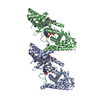
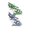
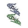
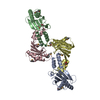
- PDB-1yj5: Molecular architecture of mammalian polynucleotide kinase, a DNA ... -
+ Open data

- Basic information
| Entry | Database: PDB / ID: 1yj5 | ||||||
|---|---|---|---|---|---|---|---|
| Title | Molecular architecture of mammalian polynucleotide kinase, a DNA repair enzyme | ||||||
 Components Components |
| ||||||
 Keywords Keywords |  TRANSFERASE / Beta sandwich / P-loop TRANSFERASE / Beta sandwich / P-loop | ||||||
| Function / homology |  Function and homology informationpolynucleotide 3'-phosphatase / polynucleotide 5'-hydroxyl-kinase / polynucleotide 3'-phosphatase activity / ATP-dependent polydeoxyribonucleotide 5'-hydroxyl-kinase activity / APEX1-Independent Resolution of AP Sites via the Single Nucleotide Replacement Pathway / DNA ligation involved in DNA repair / SCF ubiquitin ligase complex / positive regulation of double-strand break repair via nonhomologous end joining / protein K63-linked ubiquitination / positive regulation of telomere capping ...polynucleotide 3'-phosphatase / polynucleotide 5'-hydroxyl-kinase / polynucleotide 3'-phosphatase activity / ATP-dependent polydeoxyribonucleotide 5'-hydroxyl-kinase activity / APEX1-Independent Resolution of AP Sites via the Single Nucleotide Replacement Pathway / DNA ligation involved in DNA repair / SCF ubiquitin ligase complex / positive regulation of double-strand break repair via nonhomologous end joining / protein K63-linked ubiquitination / positive regulation of telomere capping / ubiquitin-like ligase-substrate adaptor activity / positive regulation of telomere maintenance via telomerase / double-strand break repair via nonhomologous end joining / site of double-strand break / double-stranded DNA binding / response to oxidative stress / phosphorylation / DNA repair / DNA damage response / nucleolus / nucleoplasm / ATP binding / nucleus Function and homology informationpolynucleotide 3'-phosphatase / polynucleotide 5'-hydroxyl-kinase / polynucleotide 3'-phosphatase activity / ATP-dependent polydeoxyribonucleotide 5'-hydroxyl-kinase activity / APEX1-Independent Resolution of AP Sites via the Single Nucleotide Replacement Pathway / DNA ligation involved in DNA repair / SCF ubiquitin ligase complex / positive regulation of double-strand break repair via nonhomologous end joining / protein K63-linked ubiquitination / positive regulation of telomere capping ...polynucleotide 3'-phosphatase / polynucleotide 5'-hydroxyl-kinase / polynucleotide 3'-phosphatase activity / ATP-dependent polydeoxyribonucleotide 5'-hydroxyl-kinase activity / APEX1-Independent Resolution of AP Sites via the Single Nucleotide Replacement Pathway / DNA ligation involved in DNA repair / SCF ubiquitin ligase complex / positive regulation of double-strand break repair via nonhomologous end joining / protein K63-linked ubiquitination / positive regulation of telomere capping / ubiquitin-like ligase-substrate adaptor activity / positive regulation of telomere maintenance via telomerase / double-strand break repair via nonhomologous end joining / site of double-strand break / double-stranded DNA binding / response to oxidative stress / phosphorylation / DNA repair / DNA damage response / nucleolus / nucleoplasm / ATP binding / nucleusSimilarity search - Function | ||||||
| Biological species |  Mus musculus (house mouse) Mus musculus (house mouse) | ||||||
| Method | X-RAY DIFFRACTION / SYNCHROTRON / MAD / Resolution: 2.8 Å | ||||||
 Authors Authors | Bernstein, N.K. / Williams, R.S. / Rakovszky, M.L. / Cui, D. / Green, R. / Karimi-Busheri, F. / Mani, R.S. / Galicia, S. / Koch, C.A. / Cass, C.E. ...Bernstein, N.K. / Williams, R.S. / Rakovszky, M.L. / Cui, D. / Green, R. / Karimi-Busheri, F. / Mani, R.S. / Galicia, S. / Koch, C.A. / Cass, C.E. / Durocher, D. / Weinfeld, M. / Glover, J.N.M. | ||||||
 Citation Citation | Journal: Mol.Cell / Year: 2005 Title: The molecular architecture of the mammalian DNA repair enzyme, polynucleotide kinase. Authors: Bernstein, N.K. / Williams, R.S. / Rakovszky, M.L. / Cui, D. / Green, R. / Karimi-Busheri, F. / Mani, R.S. / Galicia, S. / Koch, C.A. / Cass, C.E. / Durocher, D. / Weinfeld, M. / Glover, J.N. | ||||||
| History |
|
- Structure visualization
| Structure viewer | Molecule: MolmilJmol/JSmol |
|---|
- Downloads & links
-Download
| PDBx/mmCIF format | 1yj5.cif.gz | 171 KB | Display | PDBx/mmCIF format |
|---|---|---|---|---|
| PDB format | pdb1yj5.ent.gz | 142.7 KB | Display | PDB format |
| PDBx/mmJSON format | 1yj5.json.gz | Tree view | PDBx/mmJSON format | |
| Others |  Other downloads Other downloads |
-Validation report
| Arichive directory | https://data.pdbj.org/pub/pdb/validation_reports/yj/1yj5ftp://data.pdbj.org/pub/pdb/validation_reports/yj/1yj5 | HTTPS FTP |
|---|
-Related structure data


-Links
 PDBj
PDBj

- Assembly
| Deposited unit | 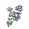
| ||||||||
|---|---|---|---|---|---|---|---|---|---|
| 1 | 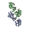
| ||||||||
| 2 | 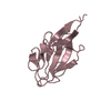
| ||||||||
| Unit cell |
| ||||||||
| Details | The biological assembly is a monomer. |
-Components
| #1: Protein | Mass: 42693.082 Da / Num. of mol.: 2 / Fragment: residues 140-522 Source method: isolated from a genetically manipulated source Source: (gene. exp.) Mus musculus (house mouse) / Plasmid: pET19b / Production host:  Escherichia coli (E. coli) / Strain (production host): BL21 gold 99 Escherichia coli (E. coli) / Strain (production host): BL21 gold 99References: UniProt: Q9JLV6, polynucleotide 5'-hydroxyl-kinase#2: Protein | | Mass: 15310.164 Da / Num. of mol.: 1 / Fragment: residues 1-143 Source method: isolated from a genetically manipulated source Source: (gene. exp.) Mus musculus (house mouse) / Plasmid: pET19b / Production host: Escherichia coli (E. coli) / Strain (production host): BL21 gold 99References: UniProt: Q9JLV6, polynucleotide 5'-hydroxyl-kinase#3: Chemical | ChemComp-SO4 / Sulfate  Mass: 96.063 Da / Num. of mol.: 12 / Source method: obtained synthetically / Formula: SO4 Mass: 96.063 Da / Num. of mol.: 12 / Source method: obtained synthetically / Formula: SO4#4: Water | ChemComp-HOH / | Water Mass: 18.015 Da / Num. of mol.: 78 / Source method: isolated from a natural source / Formula: H2O Mass: 18.015 Da / Num. of mol.: 78 / Source method: isolated from a natural source / Formula: H2O |
|---|
-Experimental details
-Experiment
| Experiment | Method: X-RAY DIFFRACTION / Number of used crystals: 1 |
|---|
- Sample preparation
| Crystal | Density Matthews: 1.8 Å3/Da / Density % sol: 31 % |
|---|---|
| Crystal grow | Temperature: 293 K / Method: vapor diffusion, sitting drop / pH: 8.3 Details: 0.1M Tris (pH 8.3 - 8.7), 18-20% PEG 5000 MME, 0.1M Li2SO4, 5mM DTT, VAPOR DIFFUSION, SITTING DROP, temperature 293K |
-Data collection
| Diffraction | Mean temperature: 100 K | ||||||||||||
|---|---|---|---|---|---|---|---|---|---|---|---|---|---|
| Diffraction source | Source: SYNCHROTRON / Site: ALS  / Beamline: 8.3.1 / Wavelength: 1.0199, 0.9793, 0.9797 / Beamline: 8.3.1 / Wavelength: 1.0199, 0.9793, 0.9797 | ||||||||||||
| Detector | Type: ADSC QUANTUM 315 / Detector: CCD / Date: Sep 20, 2003 | ||||||||||||
| Radiation | Monochromator: mirror, Si(111) double crystal / Protocol: MAD / Monochromatic (M) / Laue (L): M / Scattering type: x-ray | ||||||||||||
| Radiation wavelength |
| ||||||||||||
| Reflection | Resolution: 2.8→30 Å / Num. obs: 31529 / % possible obs: 99 % / Observed criterion σ(F): 1 / Observed criterion σ(I): 1 / Rsym value: 0.095 / Net I/σ(I): 13.1 | ||||||||||||
| Reflection shell | Resolution: 2.8→2.9 Å / Mean I/σ(I) obs: 2.4 / Rsym value: 0.448 / % possible all: 91.2 |
- Processing
| Software |
| ||||||||||||||||||||||||||||||||||||||||||||||||||||||||||||||||||||||||||||||||||||||||||||||||||||
|---|---|---|---|---|---|---|---|---|---|---|---|---|---|---|---|---|---|---|---|---|---|---|---|---|---|---|---|---|---|---|---|---|---|---|---|---|---|---|---|---|---|---|---|---|---|---|---|---|---|---|---|---|---|---|---|---|---|---|---|---|---|---|---|---|---|---|---|---|---|---|---|---|---|---|---|---|---|---|---|---|---|---|---|---|---|---|---|---|---|---|---|---|---|---|---|---|---|---|---|---|---|
| Refinement | Method to determine structure: MAD / Resolution: 2.8→29.75 Å / Cor.coef. Fo:Fc: 0.912 / Cor.coef. Fo:Fc free: 0.877 / SU B: 29.81 / SU ML: 0.28 / TLS residual ADP flag: LIKELY RESIDUAL / Cross valid method: THROUGHOUT / σ(F): 0 / ESU R: 1.033 / ESU R Free: 0.345 / Stereochemistry target values: MAXIMUM LIKELIHOOD
| ||||||||||||||||||||||||||||||||||||||||||||||||||||||||||||||||||||||||||||||||||||||||||||||||||||
| Solvent computation | Ion probe radii: 0.8 Å / Shrinkage radii: 0.8 Å / VDW probe radii: 1.2 Å / Solvent model: BABINET MODEL WITH MASK | ||||||||||||||||||||||||||||||||||||||||||||||||||||||||||||||||||||||||||||||||||||||||||||||||||||
| Displacement parameters | Biso mean: 26.374 Å2
| ||||||||||||||||||||||||||||||||||||||||||||||||||||||||||||||||||||||||||||||||||||||||||||||||||||
| Refinement step | Cycle: LAST / Resolution: 2.8→29.75 Å
| ||||||||||||||||||||||||||||||||||||||||||||||||||||||||||||||||||||||||||||||||||||||||||||||||||||
| Refine LS restraints |
| ||||||||||||||||||||||||||||||||||||||||||||||||||||||||||||||||||||||||||||||||||||||||||||||||||||
| LS refinement shell | Resolution: 2.8→2.872 Å / Total num. of bins used: 20
| ||||||||||||||||||||||||||||||||||||||||||||||||||||||||||||||||||||||||||||||||||||||||||||||||||||
| Refinement TLS params. | Method: refined / Refine-ID: X-RAY DIFFRACTION
| ||||||||||||||||||||||||||||||||||||||||||||||||||||||||||||||||||||||||||||||||||||||||||||||||||||
| Refinement TLS group |
|
