- PDB-3u16: Structure of BasE N-terminal domain from Acinetobacter baumannii ... -
+
Open data
ID or keywords:
Loading...
-
Basic information
Entry
Database: PDB / ID: 3u16
Title
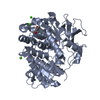
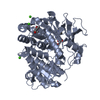
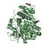
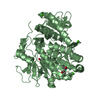
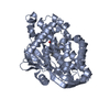
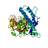
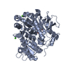
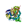
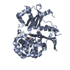
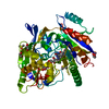
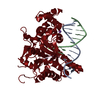
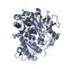
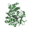
Structure of BasE N-terminal domain from Acinetobacter baumannii bound to 6-(p-benzyloxy)phenyl-1-(pyridin-4-ylmethyl)-1H-pyrazolo[3,4-b]pyridine-4-carboxylic acid.
Mass: 18.015 Da / Num. of mol.: 511 / Source method: isolated from a natural source / Formula: H2O
-
Details
Sequence details
THIS ENTRY USES A UNIPROT REFERENCE THAT IS FOR A DIFFERENT STRAIN OF A. BAUMANNI. THESE CHANGES ...THIS ENTRY USES A UNIPROT REFERENCE THAT IS FOR A DIFFERENT STRAIN OF A. BAUMANNI. THESE CHANGES ARE STRAIN RELATED DIFFERENCES AS FOUND IN GENBANK ENTRY ZP_04661818.
-
Experimental details
-
Experiment
Experiment
Method: X-RAY DIFFRACTION / Number of used crystals: 1
-
Sample preparation
Crystal
Density Matthews: 2.92 Å3/Da / Density % sol: 57.9 %
Crystal grow
Temperature: 287 K / Method: vapor diffusion, hanging drop / pH: 7.5 Details: 5-15% PEG 8000, 5% MPD, 250-600 MM CaCl2, 50 MM BTP, pH 7.5, VAPOR DIFFUSION, HANGING DROP, temperature 287K
In the structure databanks used in Yorodumi, some data are registered as the other names, "COVID-19 virus" and "2019-nCoV". Here are the details of the virus and the list of structure data.
Jan 31, 2019. EMDB accession codes are about to change! (news from PDBe EMDB page)
EMDB accession codes are about to change! (news from PDBe EMDB page)
The allocation of 4 digits for EMDB accession codes will soon come to an end. Whilst these codes will remain in use, new EMDB accession codes will include an additional digit and will expand incrementally as the available range of codes is exhausted. The current 4-digit format prefixed with “EMD-” (i.e. EMD-XXXX) will advance to a 5-digit format (i.e. EMD-XXXXX), and so on. It is currently estimated that the 4-digit codes will be depleted around Spring 2019, at which point the 5-digit format will come into force.
The EM Navigator/Yorodumi systems omit the EMD- prefix.
Related info.:Q: What is EMD? / ID/Accession-code notation in Yorodumi/EM Navigator
Yorodumi is a browser for structure data from EMDB, PDB, SASBDB, etc.
This page is also the successor to EM Navigator detail page, and also detail information page/front-end page for Omokage search.
The word "yorodu" (or yorozu) is an old Japanese word meaning "ten thousand". "mi" (miru) is to see.
Related info.:EMDB / PDB / SASBDB / Comparison of 3 databanks / Yorodumi Search / Aug 31, 2016. New EM Navigator & Yorodumi / Yorodumi Papers / Jmol/JSmol / Function and homology information / Changes in new EM Navigator and Yorodumi
 Movie
Movie Controller

 Yorodumi
 Open data
 Basic information
 Components
 Keywords
 LIGASE / ANL Superfamily / adenylating enzyme / 2 / 3-dihydroxybenzoate:Aryl Carrier protein ligase / BasF
 Function and homology information
 Authors
 Citation
 Structure visualization
 Downloads & links
 Other downloads
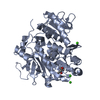


 PDBj
PDBj
 Assembly


 Mass: 436.462 Da / Num. of mol.: 2 / Source method: obtained synthetically / Formula: C26H20N4O3
 Mass: 40.078 Da / Num. of mol.: 6 / Source method: obtained synthetically / Formula: Ca
 Mass: 118.174 Da / Num. of mol.: 1 / Source method: obtained synthetically / Formula: C6H14O2 / Comment: precipitant*YM
 Mass: 118.174 Da / Num. of mol.: 1 / Source method: obtained synthetically / Formula: C6H14O2 / Comment: precipitant*YM
 Sample preparation
 / Beamline: BL11-1 / Wavelength: 0.9795 Å
 Processing