 Movie
Movie Controller

[English] 日本語
 Yorodumi
- PDB-3tzc: Crystal structure of 3-ketoacyl-(acyl-carrier-protein) reductase ... -
+ Open data

- Basic information
| Entry | Database: PDB / ID: 3tzc | ||||||
|---|---|---|---|---|---|---|---|
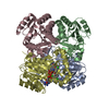
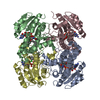
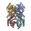
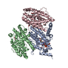
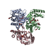
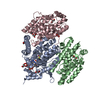
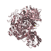
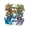
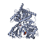
| Title | Crystal structure of 3-ketoacyl-(acyl-carrier-protein) reductase (FabG)(Y155F) from Vibrio cholerae | ||||||
 Components Components | 3-oxoacyl-[acyl-carrier protein] reductase | ||||||
 Keywords Keywords |  OXIDOREDUCTASE / Vibrio cholerae / 3-ketoacyl-(acyl-carrier-protein) reductase / FabG / Structural Genomics / Center for Structural Genomics of Infectious Diseases / CSGID OXIDOREDUCTASE / Vibrio cholerae / 3-ketoacyl-(acyl-carrier-protein) reductase / FabG / Structural Genomics / Center for Structural Genomics of Infectious Diseases / CSGID | ||||||
| Function / homology | NAD(P)-binding Rossmann-like Domain / Rossmann fold / 3-Layer(aba) Sandwich / Alpha Beta / NADP NICOTINAMIDE-ADENINE-DINUCLEOTIDE PHOSPHATE / :  Function and homology information Function and homology information | ||||||
| Biological species |   Vibrio cholerae (bacteria) Vibrio cholerae (bacteria) | ||||||
| Method | X-RAY DIFFRACTION / SYNCHROTRON / MOLECULAR REPLACEMENT / molecular replacement / Resolution: 2.45 Å | ||||||
 Authors Authors | Hou, J. / Chruszcz, M. / Zheng, H. / Grabowski, M. / Domagalski, M. / Anderson, W.F. / Minor, W. / Center for Structural Genomics of Infectious Diseases (CSGID) | ||||||
 Citation Citation | Journal: J.Bacteriol. / Year: 2015 Title: Dissecting the Structural Elements for the Activation of beta-Ketoacyl-(Acyl Carrier Protein) Reductase from Vibrio cholerae. Authors: Hou, J. / Zheng, H. / Chruszcz, M. / Zimmerman, M.D. / Shumilin, I.A. / Osinski, T. / Demas, M. / Grimshaw, S. / Minor, W. | ||||||
| History |
|
- Structure visualization
| Structure viewer | Molecule: MolmilJmol/JSmol |
|---|
- Downloads & links
-Download
| PDBx/mmCIF format | 3tzc.cif.gz | 322.2 KB | Display | PDBx/mmCIF format |
|---|---|---|---|---|
| PDB format | pdb3tzc.ent.gz | 260.9 KB | Display | PDB format |
| PDBx/mmJSON format | 3tzc.json.gz | Tree view | PDBx/mmJSON format | |
| Others |  Other downloads Other downloads |
-Validation report
| Arichive directory | https://data.pdbj.org/pub/pdb/validation_reports/tz/3tzcftp://data.pdbj.org/pub/pdb/validation_reports/tz/3tzc | HTTPS FTP |
|---|
-Related structure data
| Related structure data |  3op4C  3rroC  3rshC  3tzkC  3u09C  4i08C  4wjzC  4wk6C 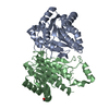 5endC C: citing same article ( |
|---|---|
| Similar structure data | |
| Other databases |

-Links
 PDBj
PDBj- Assembly
| Deposited unit | 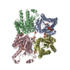
| ||||||||
|---|---|---|---|---|---|---|---|---|---|
| 1 | 
| ||||||||
| Unit cell |
|
-Components
| #1: Protein | Mass: 26342.113 Da / Num. of mol.: 4 / Mutation: Y155F Source method: isolated from a genetically manipulated source Source: (gene. exp.) Vibrio cholerae (bacteria) / Strain: MJ-1236 / Gene: VC2021, VCD_002346 / Plasmid: pMCSG7 / Production host: Escherichia coli (E. coli) / Strain (production host): BL21-CodonPlus(DE3)-RIPLReferences: UniProt: C3NP04, 3-oxoacyl-[acyl-carrier-protein] reductase #2: Chemical | ChemComp-NAP / | Nicotinamide adenine dinucleotide phosphate  Mass: 743.405 Da / Num. of mol.: 1 / Source method: obtained synthetically / Formula: C21H28N7O17P3 Mass: 743.405 Da / Num. of mol.: 1 / Source method: obtained synthetically / Formula: C21H28N7O17P3#3: Chemical | ChemComp-SO4 / Sulfate  Mass: 96.063 Da / Num. of mol.: 10 / Source method: obtained synthetically / Formula: SO4 Mass: 96.063 Da / Num. of mol.: 10 / Source method: obtained synthetically / Formula: SO4#4: Water | ChemComp-HOH / | Water Mass: 18.015 Da / Num. of mol.: 162 / Source method: isolated from a natural source / Formula: H2O Mass: 18.015 Da / Num. of mol.: 162 / Source method: isolated from a natural source / Formula: H2O |
|---|
-Experimental details
-Experiment
| Experiment | Method: X-RAY DIFFRACTION / Number of used crystals: 1 |
|---|
- Sample preparation
| Crystal | Density Matthews: 2.04 Å3/Da / Density % sol: 39.76 % |
|---|---|
| Crystal grow | Method: vapor diffusion / pH: 8.5 Details: 0.1M Tris, 2.3M Ammonium Sulfate, pH 8.5, vapor diffusion |
-Data collection
| Diffraction | Mean temperature: 100 K | |||||||||||||||||||||||||||||||||||||||||||||||||||||||||||||||||||||||||||||||||||||||||||||||||||||||||||||||||||||||||||||||||||||||||||||||||||
|---|---|---|---|---|---|---|---|---|---|---|---|---|---|---|---|---|---|---|---|---|---|---|---|---|---|---|---|---|---|---|---|---|---|---|---|---|---|---|---|---|---|---|---|---|---|---|---|---|---|---|---|---|---|---|---|---|---|---|---|---|---|---|---|---|---|---|---|---|---|---|---|---|---|---|---|---|---|---|---|---|---|---|---|---|---|---|---|---|---|---|---|---|---|---|---|---|---|---|---|---|---|---|---|---|---|---|---|---|---|---|---|---|---|---|---|---|---|---|---|---|---|---|---|---|---|---|---|---|---|---|---|---|---|---|---|---|---|---|---|---|---|---|---|---|---|---|---|---|
| Diffraction source | Source: SYNCHROTRON / Site: APS  / Beamline: 19-BM / Wavelength: 0.9792 Å / Beamline: 19-BM / Wavelength: 0.9792 Å | |||||||||||||||||||||||||||||||||||||||||||||||||||||||||||||||||||||||||||||||||||||||||||||||||||||||||||||||||||||||||||||||||||||||||||||||||||
| Detector | Type: ADSC QUANTUM 210r / Detector: CCD / Date: Jul 6, 2010 / Details: MIRRORS | |||||||||||||||||||||||||||||||||||||||||||||||||||||||||||||||||||||||||||||||||||||||||||||||||||||||||||||||||||||||||||||||||||||||||||||||||||
| Radiation | Monochromator: SI 111 CHANNEL / Protocol: SINGLE WAVELENGTH / Monochromatic (M) / Laue (L): M / Scattering type: x-ray | |||||||||||||||||||||||||||||||||||||||||||||||||||||||||||||||||||||||||||||||||||||||||||||||||||||||||||||||||||||||||||||||||||||||||||||||||||
| Radiation wavelength | Wavelength: 0.9792 Å / Relative weight: 1 | |||||||||||||||||||||||||||||||||||||||||||||||||||||||||||||||||||||||||||||||||||||||||||||||||||||||||||||||||||||||||||||||||||||||||||||||||||
| Reflection | Redundancy: 3.3 % / Av σ(I) over netI: 15.44 / Number: 100742 / Rmerge(I) obs: 0.108 / Χ2: 1.61 / D res high: 2.45 Å / D res low: 50 Å / Num. obs: 30294 / % possible obs: 98.2 | |||||||||||||||||||||||||||||||||||||||||||||||||||||||||||||||||||||||||||||||||||||||||||||||||||||||||||||||||||||||||||||||||||||||||||||||||||
| Diffraction reflection shell |
| |||||||||||||||||||||||||||||||||||||||||||||||||||||||||||||||||||||||||||||||||||||||||||||||||||||||||||||||||||||||||||||||||||||||||||||||||||
| Reflection twin |
| |||||||||||||||||||||||||||||||||||||||||||||||||||||||||||||||||||||||||||||||||||||||||||||||||||||||||||||||||||||||||||||||||||||||||||||||||||
| Reflection | Resolution: 2.45→50.01 Å / Num. obs: 30294 / % possible obs: 98.2 % / Redundancy: 3.3 % / Rmerge(I) obs: 0.108 / Χ2: 1.605 / Net I/σ(I): 7.7 | |||||||||||||||||||||||||||||||||||||||||||||||||||||||||||||||||||||||||||||||||||||||||||||||||||||||||||||||||||||||||||||||||||||||||||||||||||
| Reflection shell |
|
-Phasing
| Phasing | Method: molecular replacement |
|---|
- Processing
| Software |
| ||||||||||||||||||||||||||||||||||||||||||||||||||||||||||||
|---|---|---|---|---|---|---|---|---|---|---|---|---|---|---|---|---|---|---|---|---|---|---|---|---|---|---|---|---|---|---|---|---|---|---|---|---|---|---|---|---|---|---|---|---|---|---|---|---|---|---|---|---|---|---|---|---|---|---|---|---|---|
| Refinement | Method to determine structure: MOLECULAR REPLACEMENT / Resolution: 2.45→50 Å / Cor.coef. Fo:Fc: 0.933 / Cor.coef. Fo:Fc free: 0.897 / Occupancy max: 1 / Occupancy min: 0.3 / SU B: 10.904 / SU ML: 0.129 / Cross valid method: THROUGHOUT / σ(F): 0 / ESU R Free: 0.058 / Stereochemistry target values: MAXIMUM LIKELIHOOD Details: U VALUES WITH TLS ADDED. HYDROGENS HAVE BEEN ADDED IN THE RIDING POSITIONS.
| ||||||||||||||||||||||||||||||||||||||||||||||||||||||||||||
| Solvent computation | Ion probe radii: 0.8 Å / Shrinkage radii: 0.8 Å / VDW probe radii: 1.2 Å / Solvent model: MASK | ||||||||||||||||||||||||||||||||||||||||||||||||||||||||||||
| Displacement parameters | Biso max: 97.43 Å2 / Biso mean: 40.3658 Å2 / Biso min: 18.94 Å2
| ||||||||||||||||||||||||||||||||||||||||||||||||||||||||||||
| Refinement step | Cycle: LAST / Resolution: 2.45→50 Å
| ||||||||||||||||||||||||||||||||||||||||||||||||||||||||||||
| Refine LS restraints |
| ||||||||||||||||||||||||||||||||||||||||||||||||||||||||||||
| LS refinement shell | Resolution: 2.45→2.514 Å / Total num. of bins used: 20
| ||||||||||||||||||||||||||||||||||||||||||||||||||||||||||||
| Refinement TLS params. | Method: refined / Origin x: -1.273 Å / Origin y: 3.229 Å / Origin z: 0.935 Å
| ||||||||||||||||||||||||||||||||||||||||||||||||||||||||||||
| Refinement TLS group |
|
