 Movie
Movie Controller

[English] 日本語
 Yorodumi
- PDB-3tmp: The catalytic domain of human deubiquitinase DUBA in complex with... -
+ Open data

- Basic information
| Entry | Database: PDB / ID: 3tmp | ||||||
|---|---|---|---|---|---|---|---|
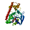
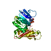
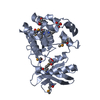
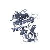
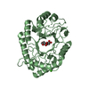
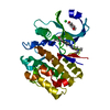
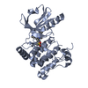
| Title | The catalytic domain of human deubiquitinase DUBA in complex with ubiquitin aldehyde | ||||||
 Components Components |
| ||||||
 Keywords Keywords | Hydrolase/Protein Binding / OTU fold /  Deubiquitinase / phosphorylation / Hydrolase-Protein Binding complex Deubiquitinase / phosphorylation / Hydrolase-Protein Binding complex | ||||||
| Function / homology |  Function and homology information Function and homology informationpositive regulation of TORC2 signaling / deubiquitinase activity / protein K48-linked deubiquitination / protein K63-linked deubiquitination / neural crest cell differentiation / K48-linked deubiquitinase activity / negative regulation of type I interferon production / K63-linked deubiquitinase activity / protein deubiquitination / Maturation of protein E ...positive regulation of TORC2 signaling / deubiquitinase activity / protein K48-linked deubiquitination / protein K63-linked deubiquitination / neural crest cell differentiation / K48-linked deubiquitinase activity / negative regulation of type I interferon production / K63-linked deubiquitinase activity / protein deubiquitination / Maturation of protein E / Maturation of protein E / ER Quality Control Compartment (ERQC) / Myoclonic epilepsy of Lafora / FLT3 signaling by CBL mutants / Prevention of phagosomal-lysosomal fusion / IRAK2 mediated activation of TAK1 complex / Alpha-protein kinase 1 signaling pathway / Glycogen synthesis / IRAK1 recruits IKK complex / IRAK1 recruits IKK complex upon TLR7/8 or 9 stimulation / Membrane binding and targetting of GAG proteins / Endosomal Sorting Complex Required For Transport (ESCRT) / IRAK2 mediated activation of TAK1 complex upon TLR7/8 or 9 stimulation / positive regulation of TORC1 signaling / PTK6 Regulates RTKs and Their Effectors AKT1 and DOK1 / Negative regulation of FLT3 / Constitutive Signaling by NOTCH1 HD Domain Mutants / Regulation of FZD by ubiquitination / TICAM1,TRAF6-dependent induction of TAK1 complex / NOTCH2 Activation and Transmission of Signal to the Nucleus / TICAM1-dependent activation of IRF3/IRF7 / APC/C:Cdc20 mediated degradation of Cyclin B / p75NTR recruits signalling complexes / Downregulation of ERBB4 signaling / TRAF6 mediated IRF7 activation in TLR7/8 or 9 signaling / APC-Cdc20 mediated degradation of Nek2A / PINK1-PRKN Mediated Mitophagy / TRAF6-mediated induction of TAK1 complex within TLR4 complex / Pexophagy / Regulation of innate immune responses to cytosolic DNA / VLDLR internalisation and degradation / InlA-mediated entry of Listeria monocytogenes into host cells / Downregulation of ERBB2:ERBB3 signaling / NF-kB is activated and signals survival / NRIF signals cell death from the nucleus / Regulation of PTEN localization / Activated NOTCH1 Transmits Signal to the Nucleus / Regulation of BACH1 activity / Translesion synthesis by REV1 / Synthesis of active ubiquitin: roles of E1 and E2 enzymes / Translesion synthesis by POLK / MAP3K8 (TPL2)-dependent MAPK1/3 activation / TICAM1, RIP1-mediated IKK complex recruitment / Downregulation of TGF-beta receptor signaling / Activation of IRF3, IRF7 mediated by TBK1, IKKε (IKBKE) / Translesion synthesis by POLI / Gap-filling DNA repair synthesis and ligation in GG-NER / Josephin domain DUBs / Regulation of activated PAK-2p34 by proteasome mediated degradation / InlB-mediated entry of Listeria monocytogenes into host cell / IKK complex recruitment mediated by RIP1 / JNK (c-Jun kinases) phosphorylation and activation mediated by activated human TAK1 / TGF-beta receptor signaling in EMT (epithelial to mesenchymal transition) / N-glycan trimming in the ER and Calnexin/Calreticulin cycle / Autodegradation of Cdh1 by Cdh1:APC/C / TNFR1-induced NF-kappa-B signaling pathway / APC/C:Cdc20 mediated degradation of Securin / Asymmetric localization of PCP proteins / SCF-beta-TrCP mediated degradation of Emi1 / TCF dependent signaling in response to WNT / NIK-->noncanonical NF-kB signaling / Regulation of NF-kappa B signaling / Ubiquitin-dependent degradation of Cyclin D / AUF1 (hnRNP D0) binds and destabilizes mRNA / TNFR2 non-canonical NF-kB pathway / activated TAK1 mediates p38 MAPK activation / Assembly of the pre-replicative complex / Vpu mediated degradation of CD4 / NOTCH3 Activation and Transmission of Signal to the Nucleus / Negative regulators of DDX58/IFIH1 signaling / Deactivation of the beta-catenin transactivating complex / Degradation of DVL / Ubiquitin Mediated Degradation of Phosphorylated Cdc25A / Regulation of signaling by CBL / Dectin-1 mediated noncanonical NF-kB signaling / Cdc20:Phospho-APC/C mediated degradation of Cyclin A / Fanconi Anemia Pathway / Hh mutants are degraded by ERAD / Negative regulation of FGFR3 signaling / Peroxisomal protein import / Recognition of DNA damage by PCNA-containing replication complex / Degradation of AXIN / Downregulation of SMAD2/3:SMAD4 transcriptional activity / Degradation of GLI1 by the proteasome / Activation of NF-kappaB in B cells / Regulation of TNFR1 signaling / Negative regulation of FGFR2 signaling / Defective CFTR causes cystic fibrosis / Termination of translesion DNA synthesis / Hedgehog ligand biogenesisSimilarity search - Function | ||||||
| Biological species |  Homo sapiens (human) Homo sapiens (human) | ||||||
| Method | X-RAY DIFFRACTION / SYNCHROTRON / MOLECULAR REPLACEMENT / Resolution: 1.91 Å | ||||||
 Authors Authors | Ma, X. / Yin, J. / Hymowitz, S. / Starovasnik, M. / Cochran, A. | ||||||
 Citation Citation | Journal: Nat.Struct.Mol.Biol. / Year: 2012 Title: Phosphorylation-dependent activity of the deubiquitinase DUBA. Authors: Huang, O.W. / Ma, X. / Yin, J. / Flinders, J. / Maurer, T. / Kayagaki, N. / Phung, Q. / Bosanac, I. / Arnott, D. / Dixit, V.M. / Hymowitz, S.G. / Starovasnik, M.A. / Cochran, A.G. | ||||||
| History |
|
- Structure visualization
| Structure viewer | Molecule: MolmilJmol/JSmol |
|---|
- Downloads & links
-Download
| PDBx/mmCIF format | 3tmp.cif.gz | 215.9 KB | Display | PDBx/mmCIF format |
|---|---|---|---|---|
| PDB format | pdb3tmp.ent.gz | 181 KB | Display | PDB format |
| PDBx/mmJSON format | 3tmp.json.gz | Tree view | PDBx/mmJSON format | |
| Others |  Other downloads Other downloads |
-Validation report
| Arichive directory | https://data.pdbj.org/pub/pdb/validation_reports/tm/3tmpftp://data.pdbj.org/pub/pdb/validation_reports/tm/3tmp | HTTPS FTP |
|---|
-Related structure data


-Links
 PDBj
PDBj


- Assembly
| Deposited unit | 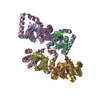
| ||||||||
|---|---|---|---|---|---|---|---|---|---|
| 1 | 
| ||||||||
| 2 | 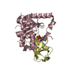
| ||||||||
| 3 | 
| ||||||||
| 4 | 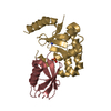
| ||||||||
| Unit cell |
|
-Components
| #1: Protein | Mass: 21256.529 Da / Num. of mol.: 4 / Fragment: catalytic or OTU domain (Residues 172-351) / Mutation: S172 phosphorylated Source method: isolated from a genetically manipulated source Source: (gene. exp.) Homo sapiens (human) / Gene: DUBA, OTUD5 / Plasmid: pET28B / Production host:  Escherichia coli (E. coli) / Strain (production host): BL21-CodonPlus (DE3)-RIL / References: UniProt: Q96G74, ubiquitinyl hydrolase 1 Escherichia coli (E. coli) / Strain (production host): BL21-CodonPlus (DE3)-RIL / References: UniProt: Q96G74, ubiquitinyl hydrolase 1#2: Protein | Mass: 8560.831 Da / Num. of mol.: 4 / Fragment: Ubiquitin-like 1 Source method: isolated from a genetically manipulated source Source: (gene. exp.) Homo sapiens (human) / Gene: UBC, ubiquitin aldehyde / Production host: Escherichia coli (E. coli) / References: UniProt: P0CG48#3: Water | ChemComp-HOH / | Water Mass: 18.015 Da / Num. of mol.: 846 / Source method: isolated from a natural source / Formula: H2O Mass: 18.015 Da / Num. of mol.: 846 / Source method: isolated from a natural source / Formula: H2O |
|---|
-Experimental details
-Experiment
| Experiment | Method: X-RAY DIFFRACTION / Number of used crystals: 1 |
|---|
- Sample preparation
| Crystal | Density Matthews: 2.11 Å3/Da / Density % sol: 41.81 % |
|---|---|
| Crystal grow | Temperature: 293 K / Method: vapor diffusion, sitting drop / pH: 5.8 Details: 20% PEG3350, 0.2 M Ammonium Fluoride, and 0.1M Bis-Tris, pH 5.8, VAPOR DIFFUSION, SITTING DROP, temperature 293K |
-Data collection
| Diffraction | Mean temperature: 100 K |
|---|---|
| Diffraction source | Source: SYNCHROTRON / Site: APS  / Beamline: 21-ID-G / Wavelength: 0.9786 Å / Beamline: 21-ID-G / Wavelength: 0.9786 Å |
| Detector | Type: MARMOSAIC 300 mm CCD / Detector: CCD / Date: Aug 16, 2010 |
| Radiation | Monochromator: C(111) / Protocol: SINGLE WAVELENGTH / Monochromatic (M) / Laue (L): M / Scattering type: x-ray |
| Radiation wavelength | Wavelength: 0.9786 Å / Relative weight: 1 |
| Reflection | Resolution: 1.91→50 Å / Num. all: 75788 / Num. obs: 73969 / % possible obs: 97.6 % / Redundancy: 2 % / Biso Wilson estimate: 22.7 Å2 / Rsym value: 0.321 / Net I/σ(I): 14.5 |
| Reflection shell | Resolution: 1.91→1.98 Å / Redundancy: 1.7 % / Mean I/σ(I) obs: 2 / Num. unique all: 7278 / Rsym value: 0.321 / % possible all: 95.4 |
- Processing
| Software |
| ||||||||||||||||||||||||||||||||||||||||||||||||||||||||||||||||||||||||||||||||||||||||||||||||||||||||||||||||||||||||||||||||||||||||||||||||||||||||||||||||||||||||||
|---|---|---|---|---|---|---|---|---|---|---|---|---|---|---|---|---|---|---|---|---|---|---|---|---|---|---|---|---|---|---|---|---|---|---|---|---|---|---|---|---|---|---|---|---|---|---|---|---|---|---|---|---|---|---|---|---|---|---|---|---|---|---|---|---|---|---|---|---|---|---|---|---|---|---|---|---|---|---|---|---|---|---|---|---|---|---|---|---|---|---|---|---|---|---|---|---|---|---|---|---|---|---|---|---|---|---|---|---|---|---|---|---|---|---|---|---|---|---|---|---|---|---|---|---|---|---|---|---|---|---|---|---|---|---|---|---|---|---|---|---|---|---|---|---|---|---|---|---|---|---|---|---|---|---|---|---|---|---|---|---|---|---|---|---|---|---|---|---|---|---|---|
| Refinement | Method to determine structure: MOLECULAR REPLACEMENT / Resolution: 1.91→46.35 Å / Cor.coef. Fo:Fc: 0.952 / Cor.coef. Fo:Fc free: 0.934 / SU B: 3.345 / SU ML: 0.099 / Cross valid method: THROUGHOUT / ESU R Free: 0.156 / Stereochemistry target values: MAXIMUM LIKELIHOOD / Details: HYDROGENS HAVE BEEN ADDED IN THE RIDING POSITIONS
| ||||||||||||||||||||||||||||||||||||||||||||||||||||||||||||||||||||||||||||||||||||||||||||||||||||||||||||||||||||||||||||||||||||||||||||||||||||||||||||||||||||||||||
| Solvent computation | Ion probe radii: 0.8 Å / Shrinkage radii: 0.8 Å / VDW probe radii: 1.4 Å / Solvent model: MASK | ||||||||||||||||||||||||||||||||||||||||||||||||||||||||||||||||||||||||||||||||||||||||||||||||||||||||||||||||||||||||||||||||||||||||||||||||||||||||||||||||||||||||||
| Displacement parameters | Biso mean: 27.269 Å2
| ||||||||||||||||||||||||||||||||||||||||||||||||||||||||||||||||||||||||||||||||||||||||||||||||||||||||||||||||||||||||||||||||||||||||||||||||||||||||||||||||||||||||||
| Refinement step | Cycle: LAST / Resolution: 1.91→46.35 Å
| ||||||||||||||||||||||||||||||||||||||||||||||||||||||||||||||||||||||||||||||||||||||||||||||||||||||||||||||||||||||||||||||||||||||||||||||||||||||||||||||||||||||||||
| Refine LS restraints |
| ||||||||||||||||||||||||||||||||||||||||||||||||||||||||||||||||||||||||||||||||||||||||||||||||||||||||||||||||||||||||||||||||||||||||||||||||||||||||||||||||||||||||||
| LS refinement shell | Resolution: 1.91→1.96 Å / Total num. of bins used: 20
|
