 Movie
Movie Controller


+ Open data

- Basic information
| Entry | Database: PDB / ID: 3sx1 | ||||||
|---|---|---|---|---|---|---|---|
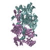
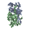
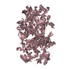
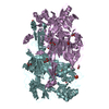
| Title | Hansenula polymorpha copper amine oxidase-1 in its apo form | ||||||
 Components Components | Peroxisomal primary amine oxidase | ||||||
 Keywords Keywords |  OXIDOREDUCTASE / peroxisome OXIDOREDUCTASE / peroxisome | ||||||
| Function / homology |  Function and homology information Function and homology information: / : / : / primary-amine oxidase / amine metabolic process / quinone binding / peroxisome / copper ion bindingSimilarity search - Function | ||||||
| Biological species |  Pichia angusta (fungus) Pichia angusta (fungus) | ||||||
| Method | X-RAY DIFFRACTION / SYNCHROTRON / MOLECULAR REPLACEMENT / Resolution: 1.73 Å | ||||||
 Authors Authors | Klema, V.J. / Johnson, B.J. / Wilmot, C.M. | ||||||
 Citation Citation | Journal: Acta Crystallogr.,Sect.F / Year: 2012 Title: The precursor form of Hansenula polymorpha copper amine oxidase 1 in complex with CuI and CoII. Authors: Klema, V.J. / Johnson, B.J. / Klinman, J.P. / Wilmot, C.M. | ||||||
| History |
|
- Structure visualization
| Structure viewer | Molecule: MolmilJmol/JSmol |
|---|
- Downloads & links
-Download
| PDBx/mmCIF format | 3sx1.cif.gz | 849.8 KB | Display | PDBx/mmCIF format |
|---|---|---|---|---|
| PDB format | pdb3sx1.ent.gz | 704.6 KB | Display | PDB format |
| PDBx/mmJSON format | 3sx1.json.gz | Tree view | PDBx/mmJSON format | |
| Others |  Other downloads Other downloads |
-Validation report
| Arichive directory | https://data.pdbj.org/pub/pdb/validation_reports/sx/3sx1ftp://data.pdbj.org/pub/pdb/validation_reports/sx/3sx1 | HTTPS FTP |
|---|
-Related structure data
| Related structure data |  3sxxC  3t0uC  2oovS S: Starting model for refinement C: citing same article ( |
|---|---|
| Similar structure data |


-Links
 PDBj
PDBj- Assembly
| Deposited unit | 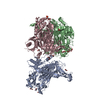
| ||||||||||||
|---|---|---|---|---|---|---|---|---|---|---|---|---|---|
| 1 | 
| ||||||||||||
| 2 | 
| ||||||||||||
| 3 | 
| ||||||||||||
| Unit cell |
| ||||||||||||
| Components on special symmetry positions |
|
-Components
| #1: Protein | Mass: 77632.617 Da / Num. of mol.: 3 Source method: isolated from a genetically manipulated source Source: (gene. exp.) Pichia angusta (fungus) / Gene: AMO / Production host:  Escherichia coli (E. coli) / References: UniProt: P12807, primary-amine oxidase Escherichia coli (E. coli) / References: UniProt: P12807, primary-amine oxidase#2: Chemical | ChemComp-GOL / Glycerol  Mass: 92.094 Da / Num. of mol.: 14 / Source method: obtained synthetically / Formula: C3H8O3 Mass: 92.094 Da / Num. of mol.: 14 / Source method: obtained synthetically / Formula: C3H8O3#3: Chemical | ChemComp-PO4 / | Phosphate  Mass: 94.971 Da / Num. of mol.: 1 / Source method: obtained synthetically / Formula: PO4 Mass: 94.971 Da / Num. of mol.: 1 / Source method: obtained synthetically / Formula: PO4#4: Water | ChemComp-HOH / | Water Mass: 18.015 Da / Num. of mol.: 2584 / Source method: isolated from a natural source / Formula: H2O Mass: 18.015 Da / Num. of mol.: 2584 / Source method: isolated from a natural source / Formula: H2O |
|---|
-Experimental details
-Experiment
| Experiment | Method: X-RAY DIFFRACTION / Number of used crystals: 1 |
|---|
- Sample preparation
| Crystal | Density Matthews: 2.57 Å3/Da / Density % sol: 52.21 % |
|---|---|
| Crystal grow | Temperature: 298 K / Method: vapor diffusion, hanging drop / pH: 6 Details: 8% PEG 8000, 0.28 M potassium phosphate, pH 6.0, VAPOR DIFFUSION, HANGING DROP, temperature 298K |
-Data collection
| Diffraction | Mean temperature: 100 K |
|---|---|
| Diffraction source | Source: SYNCHROTRON / Site: APS  / Beamline: 19-ID / Wavelength: 0.97929 Å / Beamline: 19-ID / Wavelength: 0.97929 Å |
| Detector | Type: ADSC QUANTUM 315r / Detector: CCD / Date: Nov 16, 2005 |
| Radiation | Monochromator: SAGITALLY FOCUSED Si(111) / Protocol: SINGLE WAVELENGTH / Monochromatic (M) / Laue (L): M / Scattering type: x-ray |
| Radiation wavelength | Wavelength: 0.97929 Å / Relative weight: 1 |
| Reflection | Resolution: 1.73→50 Å / Num. all: 247478 / Num. obs: 240796 / % possible obs: 97.3 % / Observed criterion σ(F): 0 / Observed criterion σ(I): -3 / Redundancy: 10 % / Rsym value: 0.074 / Net I/σ(I): 32.3 |
| Reflection shell | Resolution: 1.73→1.76 Å / Redundancy: 8.7 % / Mean I/σ(I) obs: 3.9 / Rsym value: 0.447 / % possible all: 88.1 |
- Processing
| Software |
| ||||||||||||||||||||||||||||||||||||||||||||||||||||||||||||||||||||||||||||||||||||||||||||||||||||
|---|---|---|---|---|---|---|---|---|---|---|---|---|---|---|---|---|---|---|---|---|---|---|---|---|---|---|---|---|---|---|---|---|---|---|---|---|---|---|---|---|---|---|---|---|---|---|---|---|---|---|---|---|---|---|---|---|---|---|---|---|---|---|---|---|---|---|---|---|---|---|---|---|---|---|---|---|---|---|---|---|---|---|---|---|---|---|---|---|---|---|---|---|---|---|---|---|---|---|---|---|---|
| Refinement | Method to determine structure: MOLECULAR REPLACEMENT Starting model: PDB ID 2OOV Resolution: 1.73→31.61 Å / Cor.coef. Fo:Fc: 0.973 / Cor.coef. Fo:Fc free: 0.961 / SU B: 3.133 / SU ML: 0.047 / Cross valid method: THROUGHOUT / ESU R Free: 0.082 / Stereochemistry target values: MAXIMUM LIKELIHOOD / Details: HYDROGENS HAVE BEEN ADDED IN THE RIDING POSITIONS
| ||||||||||||||||||||||||||||||||||||||||||||||||||||||||||||||||||||||||||||||||||||||||||||||||||||
| Solvent computation | Ion probe radii: 0.8 Å / Shrinkage radii: 0.8 Å / VDW probe radii: 1.4 Å / Solvent model: MASK | ||||||||||||||||||||||||||||||||||||||||||||||||||||||||||||||||||||||||||||||||||||||||||||||||||||
| Displacement parameters | Biso mean: 16.719 Å2
| ||||||||||||||||||||||||||||||||||||||||||||||||||||||||||||||||||||||||||||||||||||||||||||||||||||
| Refinement step | Cycle: LAST / Resolution: 1.73→31.61 Å
| ||||||||||||||||||||||||||||||||||||||||||||||||||||||||||||||||||||||||||||||||||||||||||||||||||||
| Refine LS restraints |
| ||||||||||||||||||||||||||||||||||||||||||||||||||||||||||||||||||||||||||||||||||||||||||||||||||||
| LS refinement shell | Resolution: 1.732→1.777 Å / Total num. of bins used: 20
| ||||||||||||||||||||||||||||||||||||||||||||||||||||||||||||||||||||||||||||||||||||||||||||||||||||
| Refinement TLS params. | Method: refined / Refine-ID: X-RAY DIFFRACTION
| ||||||||||||||||||||||||||||||||||||||||||||||||||||||||||||||||||||||||||||||||||||||||||||||||||||
| Refinement TLS group |
|
