 Movie
Movie Controller

[English] 日本語
 Yorodumi
- PDB-3nbb: Crystal structure of mutant Y305F expressed in E. coli in the cop... -
+ Open data

- Basic information
| Entry | Database: PDB / ID: 3nbb | ||||||
|---|---|---|---|---|---|---|---|
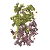
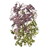
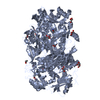
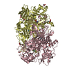
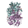
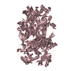
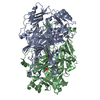
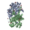
| Title | Crystal structure of mutant Y305F expressed in E. coli in the copper amine oxidase from hansenula polymorpha | ||||||
 Components Components | Peroxisomal primary amine oxidase | ||||||
 Keywords Keywords |  OXIDOREDUCTASE / amine oxidase / quinoprotein OXIDOREDUCTASE / amine oxidase / quinoprotein | ||||||
| Function / homology |  Function and homology information Function and homology information: / : / : / primary-amine oxidase / amine metabolic process / quinone binding / peroxisome / copper ion bindingSimilarity search - Function | ||||||
| Biological species |  Pichia angusta (fungus) Pichia angusta (fungus) | ||||||
| Method | X-RAY DIFFRACTION / SYNCHROTRON / MOLECULAR REPLACEMENT / Resolution: 2.05 Å | ||||||
 Authors Authors | Chen, Z. / Datta, S. / DuBois, J.L. / Klinman, J.P. / Mathews, F.S. | ||||||
 Citation Citation | Journal: Biochemistry / Year: 2010 Title: Mutation at a strictly conserved, active site tyrosine in the copper amine oxidase leads to uncontrolled oxygenase activity. Authors: Chen, Z.W. / Datta, S. / Dubois, J.L. / Klinman, J.P. / Mathews, F.S. #1: Journal: Structure / Year: 1998 Title: Copper amine oxidase from Hansenula polymorpha: the crystal structure determined at 2.4 A resolution reveals the active conformation. Authors: Li, R. / Klinman, J.P. / Mathews, F.S. | ||||||
| History |
|
- Structure visualization
| Structure viewer | Molecule: MolmilJmol/JSmol |
|---|
- Downloads & links
-Download
| PDBx/mmCIF format | 3nbb.cif.gz | 831.6 KB | Display | PDBx/mmCIF format |
|---|---|---|---|---|
| PDB format | pdb3nbb.ent.gz | 689.1 KB | Display | PDB format |
| PDBx/mmJSON format | 3nbb.json.gz | Tree view | PDBx/mmJSON format | |
| Others |  Other downloads Other downloads |
-Validation report
| Arichive directory | https://data.pdbj.org/pub/pdb/validation_reports/nb/3nbbftp://data.pdbj.org/pub/pdb/validation_reports/nb/3nbb | HTTPS FTP |
|---|
-Related structure data
| Related structure data |  3n9hC  3nbjC  1a2vS S: Starting model for refinement C: citing same article ( |
|---|---|
| Similar structure data |


-Links
 PDBj
PDBj- Assembly
| Deposited unit | 
| ||||||||
|---|---|---|---|---|---|---|---|---|---|
| 1 | 
| ||||||||
| 2 | 
| ||||||||
| 3 | 
| ||||||||
| 4 |
| ||||||||
| Unit cell |
|
-Components
| #1: Protein | Mass: 77696.617 Da / Num. of mol.: 6 / Mutation: Y305F Source method: isolated from a genetically manipulated source Source: (gene. exp.) Pichia angusta (fungus) / Gene: AMO / Plasmid: PET11A / Production host:  Escherichia coli (E. coli) / Strain (production host): BL21 / References: UniProt: P12807, primary-amine oxidase Escherichia coli (E. coli) / Strain (production host): BL21 / References: UniProt: P12807, primary-amine oxidase#2: Chemical | ChemComp-CU / Copper  Mass: 63.546 Da / Num. of mol.: 6 / Source method: obtained synthetically / Formula: Cu Mass: 63.546 Da / Num. of mol.: 6 / Source method: obtained synthetically / Formula: Cu#3: Water | ChemComp-HOH / | Water Mass: 18.015 Da / Num. of mol.: 3174 / Source method: isolated from a natural source / Formula: H2O Mass: 18.015 Da / Num. of mol.: 3174 / Source method: isolated from a natural source / Formula: H2O |
|---|
-Experimental details
-Experiment
| Experiment | Method: X-RAY DIFFRACTION / Number of used crystals: 1 |
|---|
- Sample preparation
| Crystal | Density Matthews: 2.52 Å3/Da / Density % sol: 51.13 % |
|---|---|
| Crystal grow | Temperature: 295 K / pH: 7.5 Details: 20% PEG8000, 100mM HEPES, 2% ethylene glycol, pH 7.5, VAPOR DIFFUSION, HANGING DROP, temperature 295K |
-Data collection
| Diffraction | Mean temperature: 100 K |
|---|---|
| Diffraction source | Source: SYNCHROTRON / Site: APS  / Beamline: 14-BM-C / Wavelength: 0.9 / Beamline: 14-BM-C / Wavelength: 0.9 |
| Detector | Type: ADSC QUANTUM 4 / Detector: CCD / Date: Nov 5, 2003 |
| Radiation | Monochromator: BENT GE(111) MONOCHROMATOR / Protocol: SINGLE WAVELENGTH / Monochromatic (M) / Laue (L): M / Scattering type: x-ray |
| Radiation wavelength | Wavelength: 0.9 Å / Relative weight: 1 |
| Reflection | Resolution: 2.05→40 Å / Num. obs: 217301 / % possible obs: 77.4 % / Observed criterion σ(I): -3 / Redundancy: 3.2 % / Biso Wilson estimate: 7.7 Å2 / Rmerge(I) obs: 0.067 / Net I/σ(I): 14.2 |
| Reflection shell | Resolution: 2.05→2.12 Å / Redundancy: 3.1 % / Rmerge(I) obs: 0.318 / Mean I/σ(I) obs: 3.3 |
- Processing
| Software |
| ||||||||||||||||||||||||||||||||||||||||||||||||||||||||||||
|---|---|---|---|---|---|---|---|---|---|---|---|---|---|---|---|---|---|---|---|---|---|---|---|---|---|---|---|---|---|---|---|---|---|---|---|---|---|---|---|---|---|---|---|---|---|---|---|---|---|---|---|---|---|---|---|---|---|---|---|---|---|
| Refinement | Method to determine structure: MOLECULAR REPLACEMENT Starting model: PDB ENTRY 1A2V Resolution: 2.05→36.13 Å / Rfactor Rfree error: 0.002 / Data cutoff high absF: 217092.7 / Data cutoff low absF: 0 / Isotropic thermal model: ISOTROPIC / Cross valid method: THROUGHOUT / σ(F): 0 / Stereochemistry target values: ENGH & HUBER
| ||||||||||||||||||||||||||||||||||||||||||||||||||||||||||||
| Displacement parameters | Biso mean: 29.7 Å2 | ||||||||||||||||||||||||||||||||||||||||||||||||||||||||||||
| Refine analyze |
| ||||||||||||||||||||||||||||||||||||||||||||||||||||||||||||
| Refinement step | Cycle: LAST / Resolution: 2.05→36.13 Å
| ||||||||||||||||||||||||||||||||||||||||||||||||||||||||||||
| Refine LS restraints |
| ||||||||||||||||||||||||||||||||||||||||||||||||||||||||||||
| LS refinement shell | Resolution: 2.05→2.18 Å / Rfactor Rfree error: 0.008 / Total num. of bins used: 6
| ||||||||||||||||||||||||||||||||||||||||||||||||||||||||||||
| Xplor file | Serial no: 1 / Param file: protein_rep_tpo405.par / Topol file: protein_tpo405.top |
