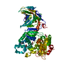
 Movie
Movie Controller


+ Open data

- Basic information
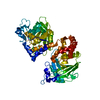
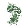
| Entry | Database: PDB / ID: 3sr9 | ||||||
|---|---|---|---|---|---|---|---|
| Title | Crystal structure of mouse PTPsigma | ||||||
 Components Components | Receptor-type tyrosine-protein phosphatase S | ||||||
 Keywords Keywords |  HYDROLASE / tyrosine phosphatase HYDROLASE / tyrosine phosphatase | ||||||
| Function / homology |  Function and homology information Function and homology informationnegative regulation of toll-like receptor 9 signaling pathway / : / : / Receptor-type tyrosine-protein phosphatases / Synaptic adhesion-like molecules / negative regulation of interferon-alpha production / : / chondroitin sulfate binding / negative regulation of collateral sprouting / negative regulation of axon regeneration ...negative regulation of toll-like receptor 9 signaling pathway / : / : / Receptor-type tyrosine-protein phosphatases / Synaptic adhesion-like molecules / negative regulation of interferon-alpha production / : / chondroitin sulfate binding / negative regulation of collateral sprouting / negative regulation of axon regeneration / establishment of endothelial intestinal barrier / negative regulation of dendritic spine development / synaptic membrane adhesion / regulation of postsynaptic density assembly / negative regulation of axon extension / corpus callosum development / negative regulation of interferon-beta production / heparan sulfate proteoglycan binding / spinal cord development / phosphoprotein phosphatase activity / regulation of presynapse assembly / peptidyl-tyrosine dephosphorylation / cerebellum development / protein dephosphorylation / protein-tyrosine-phosphatase / protein tyrosine phosphatase activity / hippocampus development / synapse organization / modulation of chemical synaptic transmission / Schaffer collateral - CA1 synapse / cerebral cortex development / negative regulation of neuron projection development / heparin binding / growth cone / perikaryon / axon / glutamatergic synapse / plasma membrane / cytosolSimilarity search - Function | ||||||
| Biological species |  Mus musculus (house mouse) Mus musculus (house mouse) | ||||||
| Method | X-RAY DIFFRACTION / SYNCHROTRON / MOLECULAR REPLACEMENT / Resolution: 2.4 Å | ||||||
 Authors Authors | Wang, J. / Hou, L. / Li, J. / Ding, J. | ||||||
 Citation Citation | Journal: Acta Biochim.Biophys.Sin. / Year: 2011 Title: Structural insights into the homology and differences between mouse protein tyrosine phosphatase-sigma and human protein tyrosine phosphatase-sigma Authors: Hou, L. / Wang, J. / Zhou, Y. / Li, J. / Zang, Y. / Li, J. | ||||||
| History |
|
- Structure visualization
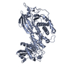
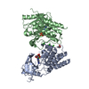
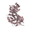
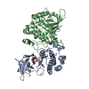
| Structure viewer | Molecule: MolmilJmol/JSmol |
|---|
- Downloads & links
-Download
| PDBx/mmCIF format | 3sr9.cif.gz | 127.7 KB | Display | PDBx/mmCIF format |
|---|---|---|---|---|
| PDB format | pdb3sr9.ent.gz | 97.4 KB | Display | PDB format |
| PDBx/mmJSON format | 3sr9.json.gz | Tree view | PDBx/mmJSON format | |
| Others |  Other downloads Other downloads |
-Validation report
| Arichive directory | https://data.pdbj.org/pub/pdb/validation_reports/sr/3sr9ftp://data.pdbj.org/pub/pdb/validation_reports/sr/3sr9 | HTTPS FTP |
|---|
-Related structure data
| Related structure data | 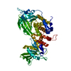 2fh7S S: Starting model for refinement |
|---|---|
| Similar structure data |


-Links
 PDBj
PDBj


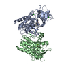
- Assembly
| Deposited unit | 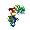
| ||||||||
|---|---|---|---|---|---|---|---|---|---|
| 1 |
| ||||||||
| Unit cell |
|
-Components
| #1: Protein | Mass: 67207.094 Da / Num. of mol.: 1 / Fragment: UNP residues 1326-1901 Source method: isolated from a genetically manipulated source Source: (gene. exp.) Mus musculus (house mouse) / Gene: Ptprs / Plasmid: pET21b / Production host:  Escherichia coli (E. coli) / Strain (production host): BL21(DE3) pLys / References: UniProt: B0V2N1, protein-tyrosine-phosphatase Escherichia coli (E. coli) / Strain (production host): BL21(DE3) pLys / References: UniProt: B0V2N1, protein-tyrosine-phosphatase |
|---|---|
| #2: Water | ChemComp-HOH / Water Mass: 18.015 Da / Num. of mol.: 89 / Source method: isolated from a natural source / Formula: H2O Mass: 18.015 Da / Num. of mol.: 89 / Source method: isolated from a natural source / Formula: H2O |
-Experimental details
-Experiment
| Experiment | Method: X-RAY DIFFRACTION / Number of used crystals: 1 |
|---|
- Sample preparation
| Crystal | Density Matthews: 2.39 Å3/Da / Density % sol: 48.53 % |
|---|---|
| Crystal grow | Temperature: 277 K / Method: vapor diffusion / pH: 7 Details: 15% polyethylene glycol 3350, 0.08M malic acid, pH 7.0, VAPOR DIFFUSION, temperature 277K |
-Data collection
| Diffraction | Mean temperature: 100 K |
|---|---|
| Diffraction source | Source: SYNCHROTRON / Site: SSRF  / Beamline: BL17U / Wavelength: 1 Å / Beamline: BL17U / Wavelength: 1 Å |
| Detector | Type: ADSC QUANTUM 315r / Detector: CCD / Date: Apr 14, 2010 |
| Radiation | Protocol: SINGLE WAVELENGTH / Monochromatic (M) / Laue (L): M / Scattering type: x-ray |
| Radiation wavelength | Wavelength: 1 Å / Relative weight: 1 |
| Reflection | Resolution: 2.4→50 Å / Num. obs: 22320 |
- Processing
| Software |
| |||||||||||||||||||||||||||||||||||||||||||||||||||||||||||||||||
|---|---|---|---|---|---|---|---|---|---|---|---|---|---|---|---|---|---|---|---|---|---|---|---|---|---|---|---|---|---|---|---|---|---|---|---|---|---|---|---|---|---|---|---|---|---|---|---|---|---|---|---|---|---|---|---|---|---|---|---|---|---|---|---|---|---|---|
| Refinement | Method to determine structure: MOLECULAR REPLACEMENT Starting model: PDB ENTRY 2FH7 Resolution: 2.4→50 Å / Cor.coef. Fo:Fc: 0.931 / Cor.coef. Fo:Fc free: 0.909 / Occupancy max: 1 / Occupancy min: 0.24 / Cross valid method: THROUGHOUT / ESU R Free: 0.323 / Stereochemistry target values: MAXIMUM LIKELIHOOD / Details: U VALUES: REFINED INDIVIDUALLY
| |||||||||||||||||||||||||||||||||||||||||||||||||||||||||||||||||
| Solvent computation | Ion probe radii: 0.8 Å / Shrinkage radii: 0.8 Å / VDW probe radii: 1.4 Å / Solvent model: MASK | |||||||||||||||||||||||||||||||||||||||||||||||||||||||||||||||||
| Displacement parameters | Biso max: 150.37 Å2 / Biso mean: 62.5058 Å2 / Biso min: 19.4 Å2
| |||||||||||||||||||||||||||||||||||||||||||||||||||||||||||||||||
| Refinement step | Cycle: LAST / Resolution: 2.4→50 Å
| |||||||||||||||||||||||||||||||||||||||||||||||||||||||||||||||||
| Refine LS restraints |
| |||||||||||||||||||||||||||||||||||||||||||||||||||||||||||||||||
| LS refinement shell | Resolution: 2.4→2.462 Å / Total num. of bins used: 20
|
