 Movie
Movie Controller


+ Open data

- Basic information
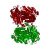
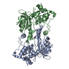
| Entry | Database: PDB / ID: 1lar | ||||||
|---|---|---|---|---|---|---|---|
| Title | CRYSTAL STRUCTURE OF THE TANDEM PHOSPHATASE DOMAINS OF RPTP LAR | ||||||
 Components Components | PROTEIN (LAR) | ||||||
 Keywords Keywords |  HYDROLASE / TYROSINE PHOSPHATEASE / LAR PROTEIN HYDROLASE / TYROSINE PHOSPHATEASE / LAR PROTEIN | ||||||
| Function / homology |  Function and homology informationchondroitin sulfate proteoglycan binding / cell surface receptor protein tyrosine phosphatase signaling pathway / neuron projection regeneration / Receptor-type tyrosine-protein phosphatases / synaptic membrane adhesion / transmembrane receptor protein tyrosine phosphatase activity / Synaptic adhesion-like molecules / regulation of axon regeneration / peptidyl-tyrosine dephosphorylation / Insulin receptor recycling ...chondroitin sulfate proteoglycan binding / cell surface receptor protein tyrosine phosphatase signaling pathway / neuron projection regeneration / Receptor-type tyrosine-protein phosphatases / synaptic membrane adhesion / transmembrane receptor protein tyrosine phosphatase activity / Synaptic adhesion-like molecules / regulation of axon regeneration / peptidyl-tyrosine dephosphorylation / Insulin receptor recycling / cell adhesion molecule binding / protein-tyrosine-phosphatase / negative regulation of receptor binding / protein tyrosine phosphatase activity / cell migration / heparin binding / cell adhesion / neuron projection / neuronal cell body / protein-containing complex binding / extracellular exosome / plasma membrane Function and homology informationchondroitin sulfate proteoglycan binding / cell surface receptor protein tyrosine phosphatase signaling pathway / neuron projection regeneration / Receptor-type tyrosine-protein phosphatases / synaptic membrane adhesion / transmembrane receptor protein tyrosine phosphatase activity / Synaptic adhesion-like molecules / regulation of axon regeneration / peptidyl-tyrosine dephosphorylation / Insulin receptor recycling ...chondroitin sulfate proteoglycan binding / cell surface receptor protein tyrosine phosphatase signaling pathway / neuron projection regeneration / Receptor-type tyrosine-protein phosphatases / synaptic membrane adhesion / transmembrane receptor protein tyrosine phosphatase activity / Synaptic adhesion-like molecules / regulation of axon regeneration / peptidyl-tyrosine dephosphorylation / Insulin receptor recycling / cell adhesion molecule binding / protein-tyrosine-phosphatase / negative regulation of receptor binding / protein tyrosine phosphatase activity / cell migration / heparin binding / cell adhesion / neuron projection / neuronal cell body / protein-containing complex binding / extracellular exosome / plasma membraneSimilarity search - Function | ||||||
| Biological species |  Homo sapiens (human) Homo sapiens (human) | ||||||
| Method | X-RAY DIFFRACTION / SYNCHROTRON / MOLECULAR REPLACEMENT / Resolution: 2 Å | ||||||
 Authors Authors | Nam, H.-J. / Poy, F. / Krueger, N. / Saito, H. / Frederick, C.A. | ||||||
 Citation Citation | Journal: Cell(Cambridge,Mass.) / Year: 1999 Title: Crystal structure of the tandem phosphatase domains of RPTP LAR. Authors: Nam, H.J. / Poy, F. / Krueger, N.X. / Saito, H. / Frederick, C.A. | ||||||
| History |
|
- Structure visualization
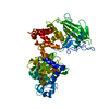
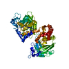
| Structure viewer | Molecule: MolmilJmol/JSmol |
|---|
- Downloads & links
-Download
| PDBx/mmCIF format | 1lar.cif.gz | 235.8 KB | Display | PDBx/mmCIF format |
|---|---|---|---|---|
| PDB format | pdb1lar.ent.gz | 188.7 KB | Display | PDB format |
| PDBx/mmJSON format | 1lar.json.gz | Tree view | PDBx/mmJSON format | |
| Others |  Other downloads Other downloads |
-Validation report
| Arichive directory | https://data.pdbj.org/pub/pdb/validation_reports/la/1larftp://data.pdbj.org/pub/pdb/validation_reports/la/1lar | HTTPS FTP |
|---|
-Related structure data
| Related structure data | 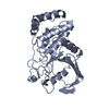 1yfoS S: Starting model for refinement |
|---|---|
| Similar structure data |


-Links
 PDBj
PDBj


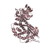
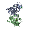
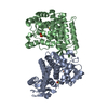
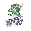
- Assembly
| Deposited unit | 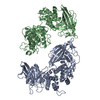
| ||||||||
|---|---|---|---|---|---|---|---|---|---|
| 1 | 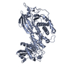
| ||||||||
| 2 | 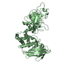
| ||||||||
| Unit cell |
| ||||||||
| Noncrystallographic symmetry (NCS) | NCS oper: (Code: given Matrix: (-0.999298, -0.034489, 0.014624), Vector : |
-Components
| #1: Protein | Mass: 66092.094 Da / Num. of mol.: 2 / Fragment: PHOSPHATASE / Mutation: P1307M Source method: isolated from a genetically manipulated source Source: (gene. exp.) Homo sapiens (human) / Species (production host): Escherichia coli / Production host:  Escherichia coli BL21(DE3) (bacteria) / Strain (production host): BL21 / Variant (production host): DE3 / References: UniProt: P10586, protein-tyrosine-phosphatase Escherichia coli BL21(DE3) (bacteria) / Strain (production host): BL21 / Variant (production host): DE3 / References: UniProt: P10586, protein-tyrosine-phosphatase#2: Water | ChemComp-HOH / | Water Mass: 18.015 Da / Num. of mol.: 473 / Source method: isolated from a natural source / Formula: H2O Mass: 18.015 Da / Num. of mol.: 473 / Source method: isolated from a natural source / Formula: H2O |
|---|
-Experimental details
-Experiment
| Experiment | Method: X-RAY DIFFRACTION / Number of used crystals: 1 |
|---|
- Sample preparation
| Crystal | Density Matthews: 2.53 Å3/Da / Density % sol: 51.45 % | ||||||||||||||||||||||||||||||
|---|---|---|---|---|---|---|---|---|---|---|---|---|---|---|---|---|---|---|---|---|---|---|---|---|---|---|---|---|---|---|---|
| Crystal grow | pH: 8 / Details: pH 8.0 | ||||||||||||||||||||||||||||||
| Crystal grow | *PLUS Method: vapor diffusion, hanging drop | ||||||||||||||||||||||||||||||
| Components of the solutions | *PLUS
|
-Data collection
| Diffraction | Mean temperature: 113 K |
|---|---|
| Diffraction source | Source: SYNCHROTRON / Site: CHESS  / Beamline: F1 / Wavelength: 0.91 / Beamline: F1 / Wavelength: 0.91 |
| Detector | Detector: CCD / Date: Dec 1, 1997 |
| Radiation | Protocol: SINGLE WAVELENGTH / Monochromatic (M) / Laue (L): M / Scattering type: x-ray |
| Radiation wavelength | Wavelength: 0.91 Å / Relative weight: 1 |
| Reflection | Resolution: 2→50 Å / Num. obs: 81312 / % possible obs: 91 % / Observed criterion σ(I): 2 / Redundancy: 3.3 % / Rmerge(I) obs: 0.064 / Rsym value: 0.064 / Net I/σ(I): 7 |
| Reflection | *PLUS Num. measured all: 267242 |
- Processing
| Software |
| ||||||||||||||||||||||||||||||||||||||||||||||||||||||||||||
|---|---|---|---|---|---|---|---|---|---|---|---|---|---|---|---|---|---|---|---|---|---|---|---|---|---|---|---|---|---|---|---|---|---|---|---|---|---|---|---|---|---|---|---|---|---|---|---|---|---|---|---|---|---|---|---|---|---|---|---|---|---|
| Refinement | Method to determine structure: MOLECULAR REPLACEMENT Starting model: PDB ENTRY 1YFO Resolution: 2→50 Å / Cross valid method: THROUGHTOUT / σ(F): 2
| ||||||||||||||||||||||||||||||||||||||||||||||||||||||||||||
| Refinement step | Cycle: LAST / Resolution: 2→50 Å
| ||||||||||||||||||||||||||||||||||||||||||||||||||||||||||||
| Refine LS restraints |
| ||||||||||||||||||||||||||||||||||||||||||||||||||||||||||||
| Software | *PLUS Name: X-PLOR / Version: 3.8 / Classification: refinement | ||||||||||||||||||||||||||||||||||||||||||||||||||||||||||||
| Refinement | *PLUS Highest resolution: 2 Å / Lowest resolution: 50 Å / σ(F): 2 / % reflection Rfree: 5 % / Rfactor obs: 0.222 | ||||||||||||||||||||||||||||||||||||||||||||||||||||||||||||
| Solvent computation | *PLUS | ||||||||||||||||||||||||||||||||||||||||||||||||||||||||||||
| Displacement parameters | *PLUS |
