 Movie
Movie Controller

[English] 日本語
 Yorodumi
- PDB-3spx: Crystal structure of O-Acetyl Serine Sulfhydrylase from Leishmani... -
+ Open data

- Basic information
| Entry | Database: PDB / ID: 3spx | ||||||
|---|---|---|---|---|---|---|---|
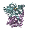
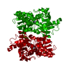
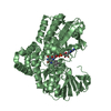
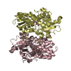
| Title | Crystal structure of O-Acetyl Serine Sulfhydrylase from Leishmania donovani | ||||||
 Components Components | O-acetyl serine sulfhydrylase | ||||||
 Keywords Keywords |  TRANSFERASE / O-Acetyl Serine Sulfhydrylase / Cysteine Synthase / Type II PLP dependent enzyme / Cysteine biocynthesis / Serine Acetyl Transferase / cytosolic TRANSFERASE / O-Acetyl Serine Sulfhydrylase / Cysteine Synthase / Type II PLP dependent enzyme / Cysteine biocynthesis / Serine Acetyl Transferase / cytosolic | ||||||
| Function / homology |  Function and homology informationcysteine synthase activity / cysteine biosynthetic process from serine / cytoplasm Function and homology informationcysteine synthase activity / cysteine biosynthetic process from serine / cytoplasmSimilarity search - Function | ||||||
| Biological species |  Leishmania donovani (eukaryote) Leishmania donovani (eukaryote) | ||||||
| Method | X-RAY DIFFRACTION / SYNCHROTRON / MOLECULAR REPLACEMENT / molecular replacement / Resolution: 1.79 Å | ||||||
 Authors Authors | Raj, I. / Gourinath, S. | ||||||
 Citation Citation | Journal: Acta Crystallogr.,Sect.D / Year: 2012 Title: The narrow active-site cleft of O-acetylserine sulfhydrylase from Leishmania donovani allows complex formation with serine acetyltransferases with a range of C-terminal sequences Authors: Raj, I. / Kumar, S. / Gourinath, S. | ||||||
| History |
|
- Structure visualization
| Structure viewer | Molecule: MolmilJmol/JSmol |
|---|
- Downloads & links
-Download
| PDBx/mmCIF format | 3spx.cif.gz | 75.6 KB | Display | PDBx/mmCIF format |
|---|---|---|---|---|
| PDB format | pdb3spx.ent.gz | 58.6 KB | Display | PDB format |
| PDBx/mmJSON format | 3spx.json.gz | Tree view | PDBx/mmJSON format | |
| Others |  Other downloads Other downloads |
-Validation report
| Arichive directory | https://data.pdbj.org/pub/pdb/validation_reports/sp/3spxftp://data.pdbj.org/pub/pdb/validation_reports/sp/3spx | HTTPS FTP |
|---|
-Related structure data


-Links
 PDBj
PDBj
- Assembly
| Deposited unit | 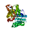
| |||||||||
|---|---|---|---|---|---|---|---|---|---|---|
| 1 | 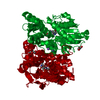
| |||||||||
| Unit cell |
| |||||||||
| Components on special symmetry positions |
|
-Components
| #1: Protein | Mass: 35907.250 Da / Num. of mol.: 1 Source method: isolated from a genetically manipulated source Source: (gene. exp.) Leishmania donovani (eukaryote) / Strain: MHOM/IM/1983/AG83 / Gene: OASS / Plasmid: pET21c / Production host:  Escherichia coli (E. coli) / Strain (production host): BL21(DE3) / References: UniProt: G1C2I2, cysteine synthase Escherichia coli (E. coli) / Strain (production host): BL21(DE3) / References: UniProt: G1C2I2, cysteine synthase |
|---|---|
| #2: Chemical | ChemComp-CL / Chloride  Mass: 35.453 Da / Num. of mol.: 1 / Source method: obtained synthetically / Formula: Cl Mass: 35.453 Da / Num. of mol.: 1 / Source method: obtained synthetically / Formula: Cl |
| #3: Chemical | ChemComp-NA /   Mass: 22.990 Da / Num. of mol.: 1 / Source method: obtained synthetically / Formula: Na Mass: 22.990 Da / Num. of mol.: 1 / Source method: obtained synthetically / Formula: Na |
| #4: Chemical | ChemComp-PEG / Diethylene glycol  Mass: 106.120 Da / Num. of mol.: 1 / Source method: obtained synthetically / Formula: C4H10O3 Mass: 106.120 Da / Num. of mol.: 1 / Source method: obtained synthetically / Formula: C4H10O3 |
| #5: Water | ChemComp-HOH / Water Mass: 18.015 Da / Num. of mol.: 196 / Source method: isolated from a natural source / Formula: H2O Mass: 18.015 Da / Num. of mol.: 196 / Source method: isolated from a natural source / Formula: H2O |
-Experimental details
-Experiment
| Experiment | Method: X-RAY DIFFRACTION / Number of used crystals: 1 |
|---|
- Sample preparation
| Crystal | Density Matthews: 2.24 Å3/Da / Density % sol: 45.02 % / Mosaicity: 0.418 ° |
|---|---|
| Crystal grow | Temperature: 289 K / Method: vapor diffusion, hanging drop / pH: 6.5 Details: KSCN, PEG 3350, Bis-tris propane, pH 6.5, VAPOR DIFFUSION, HANGING DROP, temperature 289K |
-Data collection
| Diffraction | Mean temperature: 100 K | |||||||||||||||||||||||||||||||||||||||||||||||||||||||||||||||||||||||||||||||||||||||||||||||||||||||||||||||||||||||||||||||||||||||||||||||||||
|---|---|---|---|---|---|---|---|---|---|---|---|---|---|---|---|---|---|---|---|---|---|---|---|---|---|---|---|---|---|---|---|---|---|---|---|---|---|---|---|---|---|---|---|---|---|---|---|---|---|---|---|---|---|---|---|---|---|---|---|---|---|---|---|---|---|---|---|---|---|---|---|---|---|---|---|---|---|---|---|---|---|---|---|---|---|---|---|---|---|---|---|---|---|---|---|---|---|---|---|---|---|---|---|---|---|---|---|---|---|---|---|---|---|---|---|---|---|---|---|---|---|---|---|---|---|---|---|---|---|---|---|---|---|---|---|---|---|---|---|---|---|---|---|---|---|---|---|---|
| Diffraction source | Source: SYNCHROTRON / Site: ESRF  / Beamline: BM14 / Wavelength: 0.976 Å / Beamline: BM14 / Wavelength: 0.976 Å | |||||||||||||||||||||||||||||||||||||||||||||||||||||||||||||||||||||||||||||||||||||||||||||||||||||||||||||||||||||||||||||||||||||||||||||||||||
| Detector | Type: MARMOSAIC 225 mm CCD / Detector: CCD / Date: May 1, 2010 | |||||||||||||||||||||||||||||||||||||||||||||||||||||||||||||||||||||||||||||||||||||||||||||||||||||||||||||||||||||||||||||||||||||||||||||||||||
| Radiation | Protocol: SINGLE WAVELENGTH / Monochromatic (M) / Laue (L): M / Scattering type: x-ray | |||||||||||||||||||||||||||||||||||||||||||||||||||||||||||||||||||||||||||||||||||||||||||||||||||||||||||||||||||||||||||||||||||||||||||||||||||
| Radiation wavelength | Wavelength: 0.976 Å / Relative weight: 1 | |||||||||||||||||||||||||||||||||||||||||||||||||||||||||||||||||||||||||||||||||||||||||||||||||||||||||||||||||||||||||||||||||||||||||||||||||||
| Reflection | Resolution: 1.79→50 Å / Num. all: 29852 / Num. obs: 29629 / % possible obs: 99.3 % / Redundancy: 7.8 % / Rmerge(I) obs: 0.064 / Χ2: 0.997 / Net I/σ(I): 10.9 | |||||||||||||||||||||||||||||||||||||||||||||||||||||||||||||||||||||||||||||||||||||||||||||||||||||||||||||||||||||||||||||||||||||||||||||||||||
| Reflection shell |
|
-Phasing
| Phasing | Method: molecular replacement | |||||||||
|---|---|---|---|---|---|---|---|---|---|---|
| Phasing MR |
|
- Processing
| Software |
| |||||||||||||||||||||||||||||||||||||||||||||||||||||||||||||||||
|---|---|---|---|---|---|---|---|---|---|---|---|---|---|---|---|---|---|---|---|---|---|---|---|---|---|---|---|---|---|---|---|---|---|---|---|---|---|---|---|---|---|---|---|---|---|---|---|---|---|---|---|---|---|---|---|---|---|---|---|---|---|---|---|---|---|---|
| Refinement | Method to determine structure: MOLECULAR REPLACEMENT / Resolution: 1.79→42.2 Å / Cor.coef. Fo:Fc: 0.96 / Cor.coef. Fo:Fc free: 0.944 / WRfactor Rfree: 0.207 / WRfactor Rwork: 0.167 / Occupancy max: 1 / Occupancy min: 0.5 / FOM work R set: 0.8612 / SU B: 2.406 / SU ML: 0.076 / SU R Cruickshank DPI: 0.1238 / SU Rfree: 0.1207 / Cross valid method: THROUGHOUT / ESU R Free: 0.121 / Stereochemistry target values: MAXIMUM LIKELIHOOD Details: HYDROGENS HAVE BEEN ADDED IN THE RIDING POSITIONS U VALUES: REFINED INDIVIDUALLY
| |||||||||||||||||||||||||||||||||||||||||||||||||||||||||||||||||
| Solvent computation | Ion probe radii: 0.8 Å / Shrinkage radii: 0.8 Å / VDW probe radii: 1.4 Å / Solvent model: MASK | |||||||||||||||||||||||||||||||||||||||||||||||||||||||||||||||||
| Displacement parameters | Biso max: 58.07 Å2 / Biso mean: 20.7145 Å2 / Biso min: 3.82 Å2
| |||||||||||||||||||||||||||||||||||||||||||||||||||||||||||||||||
| Refinement step | Cycle: LAST / Resolution: 1.79→42.2 Å
| |||||||||||||||||||||||||||||||||||||||||||||||||||||||||||||||||
| Refine LS restraints |
| |||||||||||||||||||||||||||||||||||||||||||||||||||||||||||||||||
| LS refinement shell | Resolution: 1.794→1.84 Å / Total num. of bins used: 20
|
