 Movie
Movie Controller


+ Open data

- Basic information
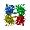
| Entry | Database: PDB / ID: 3soz | ||||||
|---|---|---|---|---|---|---|---|
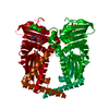
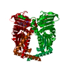
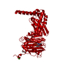
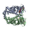
| Title | Cytoplasmic Protein STM1381 from Salmonella typhimurium LT2 | ||||||
 Components Components | Cytoplasmic Protein STM1381 Cytoplasm Cytoplasm | ||||||
 Keywords Keywords | Structural Genomics / Unknown Function / PSI-Biology / Midwest Center for Structural Genomics / MCSG / Structure and Function of Pathogen Secreted Proteins / Program for the Characterization of Secreted Effector Proteins / PCSEP | ||||||
| Function / homology | STM3548-like / Putative glutamine amidotransferase / Putative glutamine amidotransferase / Class I glutamine amidotransferase (GATase) domain / Class I glutamine amidotransferase-like / Rossmann fold / 3-Layer(aba) Sandwich / Alpha Beta / Cytoplasmic protein Function and homology information Function and homology information | ||||||
| Biological species |  Salmonella enterica subsp. enterica serovar Typhimurium (bacteria) Salmonella enterica subsp. enterica serovar Typhimurium (bacteria) | ||||||
| Method | X-RAY DIFFRACTION / SYNCHROTRON / SAD / Resolution: 2.6 Å | ||||||
 Authors Authors | Joachimiak, A. / Duke, N.E.C. / Jedrzejczak, R. / Li, H. / Adkins, J. / Brown, R. / Midwest Center for Structural Genomics (MCSG) / Program for the Characterization of Secreted Effector Proteins (PCSEP) | ||||||
 Citation Citation | Journal: To be Published Title: Cytoplasmic Protein STM1381 from Salmonella typhimurium LT2 Authors: Joachimiak, A. / Duke, N.E.C. / Jedrzejczak, R. / Li, H. / Adkins, J. / Brown, R. | ||||||
| History |
|
- Structure visualization
| Structure viewer | Molecule: MolmilJmol/JSmol |
|---|
- Downloads & links
-Download
| PDBx/mmCIF format | 3soz.cif.gz | 152.6 KB | Display | PDBx/mmCIF format |
|---|---|---|---|---|
| PDB format | pdb3soz.ent.gz | 127.8 KB | Display | PDB format |
| PDBx/mmJSON format | 3soz.json.gz | Tree view | PDBx/mmJSON format | |
| Others |  Other downloads Other downloads |
-Validation report
| Arichive directory | https://data.pdbj.org/pub/pdb/validation_reports/so/3sozftp://data.pdbj.org/pub/pdb/validation_reports/so/3soz | HTTPS FTP |
|---|
-Related structure data
| Similar structure data | |
|---|---|
| Other databases |


-Links
 PDBj
PDBj

- Assembly
| Deposited unit | 
| ||||||||
|---|---|---|---|---|---|---|---|---|---|
| 1 |
| ||||||||
| Unit cell |
| ||||||||
| Details | trimer in asymmetric unit |
-Components
| #1: Protein | Cytoplasm / ORF 245 protein Mass: 28055.889 Da / Num. of mol.: 3 Source method: isolated from a genetically manipulated source Source: (gene. exp.) Salmonella enterica subsp. enterica serovar Typhimurium (bacteria)Strain: LT2 / Gene: ORF 245, orf245, STM1381 / Plasmid: pMCSG7 / Production host: Escherichia coli (E. coli) / Strain (production host): BL21 / References: UniProt: Q9Z4S4#2: Chemical | Glycerol  Mass: 92.094 Da / Num. of mol.: 3 / Source method: obtained synthetically / Formula: C3H8O3 Mass: 92.094 Da / Num. of mol.: 3 / Source method: obtained synthetically / Formula: C3H8O3#3: Water | ChemComp-HOH / | Water Mass: 18.015 Da / Num. of mol.: 89 / Source method: isolated from a natural source / Formula: H2O Mass: 18.015 Da / Num. of mol.: 89 / Source method: isolated from a natural source / Formula: H2O |
|---|
-Experimental details
-Experiment
| Experiment | Method: X-RAY DIFFRACTION / Number of used crystals: 1 |
|---|
- Sample preparation
| Crystal | Density Matthews: 3.05 Å3/Da / Density % sol: 59.72 % |
|---|---|
| Crystal grow | Temperature: 289 K / Method: vapor diffusion, sitting drop / pH: 4.5 Details: 0.1 M Acetate pH 4.5 0.8 M NaH2PO4/1.2 M K2HPO4 (Wizard 2, screen 35), VAPOR DIFFUSION, SITTING DROP, temperature 289.0K |
-Data collection
| Diffraction | Mean temperature: 100 K |
|---|---|
| Diffraction source | Source: SYNCHROTRON / Site: APS  / Beamline: 19-ID / Wavelength: 0.9791827 Å / Beamline: 19-ID / Wavelength: 0.9791827 Å |
| Detector | Type: ADSC QUANTUM 315r / Detector: CCD / Date: Apr 3, 2011 Details: Si (111) double crystal monochromator sagitally focussing mirror |
| Radiation | Monochromator: Si (111) double crystal monochromator / Protocol: SINGLE WAVELENGTH / Monochromatic (M) / Laue (L): M / Scattering type: x-ray |
| Radiation wavelength | Wavelength: 0.9791827 Å / Relative weight: 1 |
| Reflection | Resolution: 2.6→100 Å / Num. all: 33198 / Num. obs: 33198 / Observed criterion σ(I): 3 / Redundancy: 7.6 % |
| Reflection shell | Resolution: 2.6→2.62 Å / Num. unique all: 1569 |
- Processing
| Software |
| ||||||||||||||||||||||||||||||||||||||||||||||||||||||||||||||||||||||||||||||||||||||||||||||||||||||||||||||||||||||||||||||||||||||||||||||||||||||||||||||||||||||||||
|---|---|---|---|---|---|---|---|---|---|---|---|---|---|---|---|---|---|---|---|---|---|---|---|---|---|---|---|---|---|---|---|---|---|---|---|---|---|---|---|---|---|---|---|---|---|---|---|---|---|---|---|---|---|---|---|---|---|---|---|---|---|---|---|---|---|---|---|---|---|---|---|---|---|---|---|---|---|---|---|---|---|---|---|---|---|---|---|---|---|---|---|---|---|---|---|---|---|---|---|---|---|---|---|---|---|---|---|---|---|---|---|---|---|---|---|---|---|---|---|---|---|---|---|---|---|---|---|---|---|---|---|---|---|---|---|---|---|---|---|---|---|---|---|---|---|---|---|---|---|---|---|---|---|---|---|---|---|---|---|---|---|---|---|---|---|---|---|---|---|---|---|
| Refinement | Method to determine structure: SAD / Resolution: 2.6→86.26 Å / Cor.coef. Fo:Fc: 0.949 / Cor.coef. Fo:Fc free: 0.904 / SU B: 9.941 / SU ML: 0.214 / Cross valid method: THROUGHOUT / ESU R Free: 0.304 / Stereochemistry target values: MAXIMUM LIKELIHOOD / Details: HYDROGENS HAVE BEEN ADDED IN THE RIDING POSITIONS
| ||||||||||||||||||||||||||||||||||||||||||||||||||||||||||||||||||||||||||||||||||||||||||||||||||||||||||||||||||||||||||||||||||||||||||||||||||||||||||||||||||||||||||
| Solvent computation | Ion probe radii: 0.8 Å / Shrinkage radii: 0.8 Å / VDW probe radii: 1.4 Å / Solvent model: MASK | ||||||||||||||||||||||||||||||||||||||||||||||||||||||||||||||||||||||||||||||||||||||||||||||||||||||||||||||||||||||||||||||||||||||||||||||||||||||||||||||||||||||||||
| Displacement parameters | Biso mean: 54.948 Å2
| ||||||||||||||||||||||||||||||||||||||||||||||||||||||||||||||||||||||||||||||||||||||||||||||||||||||||||||||||||||||||||||||||||||||||||||||||||||||||||||||||||||||||||
| Refinement step | Cycle: LAST / Resolution: 2.6→86.26 Å
| ||||||||||||||||||||||||||||||||||||||||||||||||||||||||||||||||||||||||||||||||||||||||||||||||||||||||||||||||||||||||||||||||||||||||||||||||||||||||||||||||||||||||||
| Refine LS restraints |
| ||||||||||||||||||||||||||||||||||||||||||||||||||||||||||||||||||||||||||||||||||||||||||||||||||||||||||||||||||||||||||||||||||||||||||||||||||||||||||||||||||||||||||
| LS refinement shell | Resolution: 2.599→2.666 Å / Total num. of bins used: 20
|
