 Movie
Movie Controller

[English] 日本語
 Yorodumi
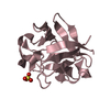
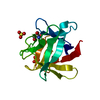
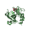
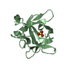
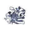
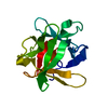
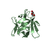
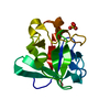
- PDB-3snv: Crystal structure of Symfoil-4T Permutation #1: de novo designed ... -
+ Open data

- Basic information
| Entry | Database: PDB / ID: 3snv | ||||||
|---|---|---|---|---|---|---|---|
| Title | Crystal structure of Symfoil-4T Permutation #1: de novo designed beta-trefoil architecture with symmetric primary structure | ||||||
 Components Components | Symfoil-4T/Permutation #1 synthetic protein | ||||||
 Keywords Keywords |  DE NOVO PROTEIN / beta-trefoil DE NOVO PROTEIN / beta-trefoil | ||||||
| Function / homology | Trefoil (Acidic Fibroblast Growth Factor, subunit A) - #50 / Trefoil (Acidic Fibroblast Growth Factor, subunit A) / Trefoil / Mainly Beta Function and homology information Function and homology information | ||||||
| Biological species |  Homo sapiens (human) Homo sapiens (human) | ||||||
| Method | X-RAY DIFFRACTION / MOLECULAR REPLACEMENT / Resolution: 2.2 Å | ||||||
 Authors Authors | Blaber, M. / Longo, L. / Lee, J. | ||||||
 Citation Citation | Journal: To be Published Title: Folding pathway redundancy in symmetric protein architecture Authors: Longo, L. / Lee, J. / Blaber, M. | ||||||
| History |
|
- Structure visualization
| Structure viewer | Molecule: MolmilJmol/JSmol |
|---|
- Downloads & links
-Download
| PDBx/mmCIF format | 3snv.cif.gz | 63.3 KB | Display | PDBx/mmCIF format |
|---|---|---|---|---|
| PDB format | pdb3snv.ent.gz | 46.6 KB | Display | PDB format |
| PDBx/mmJSON format | 3snv.json.gz | Tree view | PDBx/mmJSON format | |
| Others |  Other downloads Other downloads |
-Validation report
| Arichive directory | https://data.pdbj.org/pub/pdb/validation_reports/sn/3snvftp://data.pdbj.org/pub/pdb/validation_reports/sn/3snv | HTTPS FTP |
|---|
-Related structure data
| Related structure data |  3o4bS S: Starting model for refinement |
|---|---|
| Similar structure data |


-Links
 PDBj
PDBj
- Assembly
| Deposited unit | 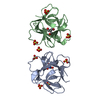
| ||||||||
|---|---|---|---|---|---|---|---|---|---|
| 1 | 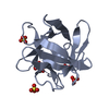
| ||||||||
| 2 | 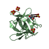
| ||||||||
| Unit cell |
|
-Components
| #1: Protein | Mass: 15994.363 Da / Num. of mol.: 2 Source method: isolated from a genetically manipulated source Source: (gene. exp.) Homo sapiens (human) / Production host:  Escherichia coli (E. coli) Escherichia coli (E. coli)#2: Chemical | Tris  Mass: 122.143 Da / Num. of mol.: 2 / Source method: obtained synthetically / Formula: C4H12NO3 / Comment: pH buffer*YM Mass: 122.143 Da / Num. of mol.: 2 / Source method: obtained synthetically / Formula: C4H12NO3 / Comment: pH buffer*YM#3: Chemical | ChemComp-SO4 / Sulfate  Mass: 96.063 Da / Num. of mol.: 8 / Source method: obtained synthetically / Formula: SO4 Mass: 96.063 Da / Num. of mol.: 8 / Source method: obtained synthetically / Formula: SO4#4: Water | ChemComp-HOH / | Water Mass: 18.015 Da / Num. of mol.: 72 / Source method: isolated from a natural source / Formula: H2O Mass: 18.015 Da / Num. of mol.: 72 / Source method: isolated from a natural source / Formula: H2OSequence details | THIS IS A CIRCULAR PERMUTATION OF THE SYMFOIL-4T PROTEIN (PDB ACCESSION 3O4B). IN THIS PERMUTATION ...THIS IS A CIRCULAR PERMUTATIO | |
|---|
-Experimental details
-Experiment
| Experiment | Method: X-RAY DIFFRACTION / Number of used crystals: 1 |
|---|
- Sample preparation
| Crystal | Density Matthews: 2.04 Å3/Da / Density % sol: 39.57 % |
|---|---|
| Crystal grow | Temperature: 298 K / Method: vapor diffusion, hanging drop / pH: 7 Details: 1.9 M ammonium sulfate, 0.2 M lithium sulfate, pH 7.0, VAPOR DIFFUSION, HANGING DROP, temperature 298K |
-Data collection
| Diffraction | Mean temperature: 100 K |
|---|---|
| Diffraction source | Source: ROTATING ANODE / Type: RIGAKU RUH2R / Wavelength: 1.5418 Å |
| Detector | Type: MAR CCD 165 mm / Detector: CCD / Date: Dec 19, 2010 / Details: Osmic confocal |
| Radiation | Monochromator: Osmic confocal / Protocol: SINGLE WAVELENGTH / Monochromatic (M) / Laue (L): M / Scattering type: x-ray |
| Radiation wavelength | Wavelength: 1.5418 Å / Relative weight: 1 |
| Reflection | Resolution: 2.2→40 Å / Num. all: 13099 / Num. obs: 12051 / % possible obs: 92 % / Observed criterion σ(F): 1 / Observed criterion σ(I): 1 |
| Reflection shell | Resolution: 2.2→2.26 Å / Redundancy: 2.2 % / Rmerge(I) obs: 0.331 / Mean I/σ(I) obs: 4.64 / Num. unique all: 636 / % possible all: 57.9 |
- Processing
| Software |
| |||||||||||||||||||||||||||||||||||
|---|---|---|---|---|---|---|---|---|---|---|---|---|---|---|---|---|---|---|---|---|---|---|---|---|---|---|---|---|---|---|---|---|---|---|---|---|
| Refinement | Method to determine structure: MOLECULAR REPLACEMENT Starting model: PDB entry 3O4B Resolution: 2.2→23.226 Å / Occupancy max: 1 / Occupancy min: 0 / FOM work R set: 0.7957 / SU ML: 0.39 / σ(F): 2 / Phase error: 27.35 / Stereochemistry target values: ML
| |||||||||||||||||||||||||||||||||||
| Solvent computation | Shrinkage radii: 0.61 Å / VDW probe radii: 0.9 Å / Solvent model: FLAT BULK SOLVENT MODEL / Bsol: 30.747 Å2 / ksol: 0.377 e/Å3 | |||||||||||||||||||||||||||||||||||
| Displacement parameters | Biso max: 95.08 Å2 / Biso mean: 40.2462 Å2 / Biso min: 15.04 Å2
| |||||||||||||||||||||||||||||||||||
| Refinement step | Cycle: LAST / Resolution: 2.2→23.226 Å
| |||||||||||||||||||||||||||||||||||
| Refine LS restraints |
| |||||||||||||||||||||||||||||||||||
| LS refinement shell | Refine-ID: X-RAY DIFFRACTION / Total num. of bins used: 4
|
