 Movie
Movie Controller

[English] 日本語
 Yorodumi
- PDB-3sl1: Crystal Structure of P. falciparum arginase complexed with 2-amin... -
+ Open data

- Basic information
| Entry | Database: PDB / ID: 3sl1 | ||||||
|---|---|---|---|---|---|---|---|
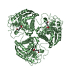
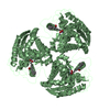
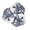
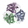
| Title | Crystal Structure of P. falciparum arginase complexed with 2-amino-6-borono-2-methylhexanoic acid | ||||||
 Components Components | Arginase | ||||||
 Keywords Keywords | HYDROLASE/HYDROLASE INHIBITOR / metallohydrolase / arginase fold / HYDROLASE-HYDROLASE INHIBITOR complex | ||||||
| Function / homology |  Function and homology informationUrea cycle / Neutrophil degranulation / arginase / arginine catabolic process to ornithine / arginase activity / urea cycle / manganese ion binding / identical protein binding / nucleus / cytosol / cytoplasm Function and homology informationUrea cycle / Neutrophil degranulation / arginase / arginine catabolic process to ornithine / arginase activity / urea cycle / manganese ion binding / identical protein binding / nucleus / cytosol / cytoplasmSimilarity search - Function | ||||||
| Biological species |  Plasmodium falciparum (malaria parasite P. falciparum) Plasmodium falciparum (malaria parasite P. falciparum) | ||||||
| Method | X-RAY DIFFRACTION / SYNCHROTRON / MOLECULAR REPLACEMENT / Resolution: 1.902 Å | ||||||
 Authors Authors | Dowling, D.P. / Ilies, M. / Christianson, D.W. | ||||||
 Citation Citation | Journal: J.Med.Chem. / Year: 2011 Title: Binding of alpha , alpha-disubstituted amino acids to arginase suggests new avenues for inhibitor design. Authors: Ilies, M. / Di Costanzo, L. / Dowling, D.P. / Thorn, K.J. / Christianson, D.W. | ||||||
| History |
|
- Structure visualization
| Structure viewer | Molecule: MolmilJmol/JSmol |
|---|
- Downloads & links
-Download
| PDBx/mmCIF format | 3sl1.cif.gz | 144.7 KB | Display | PDBx/mmCIF format |
|---|---|---|---|---|
| PDB format | pdb3sl1.ent.gz | 112.2 KB | Display | PDB format |
| PDBx/mmJSON format | 3sl1.json.gz | Tree view | PDBx/mmJSON format | |
| Others |  Other downloads Other downloads |
-Validation report
| Arichive directory | https://data.pdbj.org/pub/pdb/validation_reports/sl/3sl1ftp://data.pdbj.org/pub/pdb/validation_reports/sl/3sl1 | HTTPS FTP |
|---|
-Related structure data
| Related structure data |  3gmzC  3gn0C  3sjtC  3skkC  3sl0C  3mmrS S: Starting model for refinement C: citing same article ( |
|---|---|
| Similar structure data |


-Links
 PDBj
PDBj- Assembly
| Deposited unit | 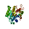
| ||||||||||||
|---|---|---|---|---|---|---|---|---|---|---|---|---|---|
| 1 | 
| ||||||||||||
| 2 | x 6
| ||||||||||||
| Unit cell |
| ||||||||||||
| Components on special symmetry positions |
|
-Components
| #1: Protein | Mass: 46452.039 Da / Num. of mol.: 1 / Fragment: UNP residues 22-411 Source method: isolated from a genetically manipulated source Source: (gene. exp.) Plasmodium falciparum (malaria parasite P. falciparum)Strain: 3D7 / Gene: pfi0320w / Plasmid: pET28a / Production host:  Escherichia coli (E. coli) / Strain (production host): Bl21 Codonplustm(de3) Ril / References: UniProt: Q8I384, arginase Escherichia coli (E. coli) / Strain (production host): Bl21 Codonplustm(de3) Ril / References: UniProt: Q8I384, arginase | ||||
|---|---|---|---|---|---|
| #2: Chemical |   Mass: 54.938 Da / Num. of mol.: 2 / Source method: obtained synthetically / Formula: Mn Mass: 54.938 Da / Num. of mol.: 2 / Source method: obtained synthetically / Formula: Mn#3: Chemical | ChemComp-FB6 / |   Type: L-peptide linking / Mass: 189.017 Da / Num. of mol.: 1 / Source method: obtained synthetically / Formula: C7H16BNO4 Type: L-peptide linking / Mass: 189.017 Da / Num. of mol.: 1 / Source method: obtained synthetically / Formula: C7H16BNO4#4: Water | ChemComp-HOH / | Water Mass: 18.015 Da / Num. of mol.: 212 / Source method: isolated from a natural source / Formula: H2O Mass: 18.015 Da / Num. of mol.: 212 / Source method: isolated from a natural source / Formula: H2O |
-Experimental details
-Experiment
| Experiment | Method: X-RAY DIFFRACTION / Number of used crystals: 3 |
|---|
- Sample preparation
| Crystal | Density Matthews: 3.05 Å3/Da / Density % sol: 59.63 % |
|---|---|
| Crystal grow | Temperature: 294 K / Method: vapor diffusion, sitting drop / pH: 8 Details: 1.2 M Na/K Phosphate (pH 8.0), VAPOR DIFFUSION, SITTING DROP, temperature 294K |
-Data collection
| Diffraction |
| ||||||||||||||||||||
|---|---|---|---|---|---|---|---|---|---|---|---|---|---|---|---|---|---|---|---|---|---|
| Diffraction source |
| ||||||||||||||||||||
| Detector |
| ||||||||||||||||||||
| Radiation |
| ||||||||||||||||||||
| Radiation wavelength | Wavelength: 0.97918 Å / Relative weight: 1 | ||||||||||||||||||||
| Reflection | Resolution: 1.9→50 Å / Num. obs: 44684 / % possible obs: 99.9 % / Redundancy: 9.1 % / Rmerge(I) obs: 0.09 / Net I/σ(I): 23.6 | ||||||||||||||||||||
| Reflection shell | Resolution: 1.9→1.97 Å / Redundancy: 4.4 % / Rmerge(I) obs: 0.444 / Mean I/σ(I) obs: 3.4 / Num. unique all: 4393 / % possible all: 99.2 |

- Processing
| Software |
| |||||||||||||||||||||||||||||||||||||||||||||||||||||||||||||||||||||||||||||||||||||||||||||||||||||||||||||||||||||||||||||||||||||||||||||||||||||||||||||||||||||||||||||||
|---|---|---|---|---|---|---|---|---|---|---|---|---|---|---|---|---|---|---|---|---|---|---|---|---|---|---|---|---|---|---|---|---|---|---|---|---|---|---|---|---|---|---|---|---|---|---|---|---|---|---|---|---|---|---|---|---|---|---|---|---|---|---|---|---|---|---|---|---|---|---|---|---|---|---|---|---|---|---|---|---|---|---|---|---|---|---|---|---|---|---|---|---|---|---|---|---|---|---|---|---|---|---|---|---|---|---|---|---|---|---|---|---|---|---|---|---|---|---|---|---|---|---|---|---|---|---|---|---|---|---|---|---|---|---|---|---|---|---|---|---|---|---|---|---|---|---|---|---|---|---|---|---|---|---|---|---|---|---|---|---|---|---|---|---|---|---|---|---|---|---|---|---|---|---|---|---|
| Refinement | Method to determine structure: MOLECULAR REPLACEMENT Starting model: 3MMR Resolution: 1.902→37.273 Å / SU ML: 0.45 / Isotropic thermal model: 31.3 / Cross valid method: THROUGHOUT / σ(F): 1.34 / Phase error: 16.82 / Stereochemistry target values: ML
| |||||||||||||||||||||||||||||||||||||||||||||||||||||||||||||||||||||||||||||||||||||||||||||||||||||||||||||||||||||||||||||||||||||||||||||||||||||||||||||||||||||||||||||||
| Solvent computation | Shrinkage radii: 1.17 Å / VDW probe radii: 1.4 Å / Solvent model: FLAT BULK SOLVENT MODEL / Bsol: 40.535 Å2 / ksol: 0.327 e/Å3 | |||||||||||||||||||||||||||||||||||||||||||||||||||||||||||||||||||||||||||||||||||||||||||||||||||||||||||||||||||||||||||||||||||||||||||||||||||||||||||||||||||||||||||||||
| Displacement parameters |
| |||||||||||||||||||||||||||||||||||||||||||||||||||||||||||||||||||||||||||||||||||||||||||||||||||||||||||||||||||||||||||||||||||||||||||||||||||||||||||||||||||||||||||||||
| Refinement step | Cycle: LAST / Resolution: 1.902→37.273 Å
| |||||||||||||||||||||||||||||||||||||||||||||||||||||||||||||||||||||||||||||||||||||||||||||||||||||||||||||||||||||||||||||||||||||||||||||||||||||||||||||||||||||||||||||||
| Refine LS restraints |
| |||||||||||||||||||||||||||||||||||||||||||||||||||||||||||||||||||||||||||||||||||||||||||||||||||||||||||||||||||||||||||||||||||||||||||||||||||||||||||||||||||||||||||||||
| LS refinement shell |
| |||||||||||||||||||||||||||||||||||||||||||||||||||||||||||||||||||||||||||||||||||||||||||||||||||||||||||||||||||||||||||||||||||||||||||||||||||||||||||||||||||||||||||||||
| Refinement TLS params. | Method: refined / Refine-ID: X-RAY DIFFRACTION
| |||||||||||||||||||||||||||||||||||||||||||||||||||||||||||||||||||||||||||||||||||||||||||||||||||||||||||||||||||||||||||||||||||||||||||||||||||||||||||||||||||||||||||||||
| Refinement TLS group |
|
