 Movie
Movie Controller

[English] 日本語
 Yorodumi
- PDB-3sfm: Novel crystallization conditions for tandem variant R67 DHFR yiel... -
+ Open data

- Basic information
| Entry | Database: PDB / ID: 3sfm | ||||||
|---|---|---|---|---|---|---|---|
| Title | Novel crystallization conditions for tandem variant R67 DHFR yields wild-type crystal structure | ||||||
 Components Components | Dihydrofolate reductase type 2 | ||||||
 Keywords Keywords |  OXIDOREDUCTASE / dihydrofolate reductase / antibiotic resistance / in situ proteolysis / type II DHFR / SH3 / reductase / DHF and NADPH-binding OXIDOREDUCTASE / dihydrofolate reductase / antibiotic resistance / in situ proteolysis / type II DHFR / SH3 / reductase / DHF and NADPH-binding | ||||||
| Function / homology |  Function and homology information Function and homology informationresponse to methotrexate / dihydrofolate reductase / dihydrofolate reductase activity / tetrahydrofolate biosynthetic process / one-carbon metabolic process / response to xenobiotic stimulus / response to antibioticSimilarity search - Function | ||||||
| Biological species |  Escherichia coli (E. coli) Escherichia coli (E. coli) | ||||||
| Method | X-RAY DIFFRACTION / MOLECULAR REPLACEMENT / Resolution: 1.4 Å | ||||||
 Authors Authors | Yachnin, B.J. / Berghuis, A.M. | ||||||
 Citation Citation | Journal: Acta Crystallogr.,Sect.F / Year: 2011 Title: Novel crystallization conditions for tandem variant R67 DHFR yield a wild-type crystal structure. Authors: Yachnin, B.J. / Colin, D.Y. / Volpato, J.P. / Ebert, M. / Pelletier, J.N. / Berghuis, A.M. | ||||||
| History |
|
- Structure visualization
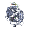
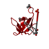
| Structure viewer | Molecule: MolmilJmol/JSmol |
|---|
- Downloads & links
-Download
| PDBx/mmCIF format | 3sfm.cif.gz | 41.4 KB | Display | PDBx/mmCIF format |
|---|---|---|---|---|
| PDB format | pdb3sfm.ent.gz | 28.3 KB | Display | PDB format |
| PDBx/mmJSON format | 3sfm.json.gz | Tree view | PDBx/mmJSON format | |
| Others |  Other downloads Other downloads |
-Validation report
| Arichive directory | https://data.pdbj.org/pub/pdb/validation_reports/sf/3sfmftp://data.pdbj.org/pub/pdb/validation_reports/sf/3sfm | HTTPS FTP |
|---|
-Related structure data
| Related structure data |  2rh2S S: Starting model for refinement |
|---|---|
| Similar structure data |


-Links
 PDBj
PDBj


- Assembly
| Deposited unit | 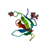
| ||||||||
|---|---|---|---|---|---|---|---|---|---|
| 1 | 
| ||||||||
| Unit cell |
|
-Components
| #1: Protein | Mass: 6733.513 Da / Num. of mol.: 1 / Fragment: Residues 21-78 Source method: isolated from a genetically manipulated source Source: (gene. exp.) Escherichia coli (E. coli) / Gene: dfrB / Plasmid: pQE-32-derived / Production host: Escherichia coli (E. coli) / Strain (production host): BL21(pRep4) / References: UniProt: P00383, dihydrofolate reductase | ||||
|---|---|---|---|---|---|
| #2: Chemical | 2-Methyl-2,4-pentanediol  Mass: 118.174 Da / Num. of mol.: 3 / Source method: obtained synthetically / Formula: C6H14O2 / Comment: precipitant*YM Mass: 118.174 Da / Num. of mol.: 3 / Source method: obtained synthetically / Formula: C6H14O2 / Comment: precipitant*YM#3: Water | ChemComp-HOH / | Water Mass: 18.015 Da / Num. of mol.: 59 / Source method: isolated from a natural source / Formula: H2O Mass: 18.015 Da / Num. of mol.: 59 / Source method: isolated from a natural source / Formula: H2OSequence details | THE FULL LENGTH OF THE EXPRESSED PROTEIN (TOTAL OF 171 residues: ...THE FULL LENGTH OF THE EXPRESSED PROTEIN (TOTAL OF 171 residues: MRGSHHHHHH | |
-Experimental details
-Experiment
| Experiment | Method: X-RAY DIFFRACTION / Number of used crystals: 1 |
|---|
- Sample preparation
| Crystal | Density Matthews: 2.35 Å3/Da / Density % sol: 47.7 % |
|---|---|
| Crystal grow | Temperature: 277 K / Method: vapor diffusion, hanging drop / pH: 7.2 Details: 0.1 M sodium phosphate, 55% MPD, pH 7.2, VAPOR DIFFUSION, HANGING DROP, temperature 277K |
-Data collection
| Diffraction | Mean temperature: 93 K |
|---|---|
| Diffraction source | Source: ROTATING ANODE / Type: RIGAKU RUH3R / Wavelength: 1.5418 Å |
| Detector | Type: RIGAKU RAXIS IV++ / Detector: IMAGE PLATE / Date: Sep 16, 2009 / Details: Osmic Confocal Blue |
| Radiation | Monochromator: Rotating copper anode (1.5418) / Protocol: SINGLE WAVELENGTH / Monochromatic (M) / Laue (L): M / Scattering type: x-ray |
| Radiation wavelength | Wavelength: 1.5418 Å / Relative weight: 1 |
| Reflection | Resolution: 1.4→50 Å / Num. all: 12169 / Num. obs: 12169 / % possible obs: 100 % / Redundancy: 23.8 % / Biso Wilson estimate: 16.788 Å2 / Rmerge(I) obs: 0.061 / Rsym value: 0.061 / Net I/σ(I): 18.5 |
| Reflection shell | Resolution: 1.4→1.43 Å / Redundancy: 13.9 % / Rmerge(I) obs: 0.371 / Num. unique all: 802 / Rsym value: 0.371 / % possible all: 100 |
- Processing
| Software |
| ||||||||||||||||||||||||||||||||||||||||||||||||||||||||||||||||||||||||||||||||||||||||||||||||||||||||||||||||||||||||||||||||||||||||||||||||||||||||||||||||||||||||||
|---|---|---|---|---|---|---|---|---|---|---|---|---|---|---|---|---|---|---|---|---|---|---|---|---|---|---|---|---|---|---|---|---|---|---|---|---|---|---|---|---|---|---|---|---|---|---|---|---|---|---|---|---|---|---|---|---|---|---|---|---|---|---|---|---|---|---|---|---|---|---|---|---|---|---|---|---|---|---|---|---|---|---|---|---|---|---|---|---|---|---|---|---|---|---|---|---|---|---|---|---|---|---|---|---|---|---|---|---|---|---|---|---|---|---|---|---|---|---|---|---|---|---|---|---|---|---|---|---|---|---|---|---|---|---|---|---|---|---|---|---|---|---|---|---|---|---|---|---|---|---|---|---|---|---|---|---|---|---|---|---|---|---|---|---|---|---|---|---|---|---|---|
| Refinement | Method to determine structure: MOLECULAR REPLACEMENT Starting model: PDB Entry 2RH2 Resolution: 1.4→23.97 Å / Cor.coef. Fo:Fc: 0.971 / Cor.coef. Fo:Fc free: 0.979 / SU B: 1.218 / SU ML: 0.023 / Isotropic thermal model: Anisotropic / Cross valid method: THROUGHOUT / ESU R Free: 0.052 / Stereochemistry target values: MAXIMUM LIKELIHOOD
| ||||||||||||||||||||||||||||||||||||||||||||||||||||||||||||||||||||||||||||||||||||||||||||||||||||||||||||||||||||||||||||||||||||||||||||||||||||||||||||||||||||||||||
| Solvent computation | Ion probe radii: 0.8 Å / Shrinkage radii: 0.8 Å / VDW probe radii: 1.4 Å / Solvent model: MASK | ||||||||||||||||||||||||||||||||||||||||||||||||||||||||||||||||||||||||||||||||||||||||||||||||||||||||||||||||||||||||||||||||||||||||||||||||||||||||||||||||||||||||||
| Displacement parameters | Biso mean: 17.816 Å2
| ||||||||||||||||||||||||||||||||||||||||||||||||||||||||||||||||||||||||||||||||||||||||||||||||||||||||||||||||||||||||||||||||||||||||||||||||||||||||||||||||||||||||||
| Refinement step | Cycle: LAST / Resolution: 1.4→23.97 Å
| ||||||||||||||||||||||||||||||||||||||||||||||||||||||||||||||||||||||||||||||||||||||||||||||||||||||||||||||||||||||||||||||||||||||||||||||||||||||||||||||||||||||||||
| Refine LS restraints |
| ||||||||||||||||||||||||||||||||||||||||||||||||||||||||||||||||||||||||||||||||||||||||||||||||||||||||||||||||||||||||||||||||||||||||||||||||||||||||||||||||||||||||||
| LS refinement shell | Resolution: 1.4→1.435 Å / Total num. of bins used: 20
|
