 Movie
Movie Controller

[English] 日本語
 Yorodumi
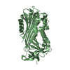
- PDB-3s2u: Crystal structure of the Pseudomonas aeruginosa MurG:UDP-GlcNAc s... -
+ Open data

- Basic information
| Entry | Database: PDB / ID: 3s2u | ||||||
|---|---|---|---|---|---|---|---|
| Title | Crystal structure of the Pseudomonas aeruginosa MurG:UDP-GlcNAc substrate complex | ||||||
 Components Components | UDP-N-acetylglucosamine--N-acetylmuramyl-(pentapeptide) pyrophosphoryl-undecaprenol N-acetylglucosamine transferase | ||||||
 Keywords Keywords |  TRANSFERASE / N-acetylglucosaminyl transferase TRANSFERASE / N-acetylglucosaminyl transferase | ||||||
| Function / homology |  Function and homology informationundecaprenyldiphospho-muramoylpentapeptide beta-N-acetylglucosaminyltransferase / undecaprenyldiphospho-muramoylpentapeptide beta-N-acetylglucosaminyltransferase activity / UDP-N-acetyl-D-glucosamine:N-acetylmuramoyl-L-alanyl-D-glutamyl-meso-2,6-diaminopimelyl-D-alanyl-D-alanine-diphosphoundecaprenol 4-beta-N-acetylglucosaminlytransferase activity / lipid glycosylation / peptidoglycan biosynthetic process / cell wall organization / regulation of cell shape / carbohydrate metabolic process / cell cycle / cell division / plasma membrane Function and homology informationundecaprenyldiphospho-muramoylpentapeptide beta-N-acetylglucosaminyltransferase / undecaprenyldiphospho-muramoylpentapeptide beta-N-acetylglucosaminyltransferase activity / UDP-N-acetyl-D-glucosamine:N-acetylmuramoyl-L-alanyl-D-glutamyl-meso-2,6-diaminopimelyl-D-alanyl-D-alanine-diphosphoundecaprenol 4-beta-N-acetylglucosaminlytransferase activity / lipid glycosylation / peptidoglycan biosynthetic process / cell wall organization / regulation of cell shape / carbohydrate metabolic process / cell cycle / cell division / plasma membraneSimilarity search - Function | ||||||
| Biological species |   Pseudomonas aeruginosa (bacteria) Pseudomonas aeruginosa (bacteria) | ||||||
| Method | X-RAY DIFFRACTION / SYNCHROTRON / Resolution: 2.23 Å | ||||||
 Authors Authors | Brown, K. / Vial, S.C.M. / Dedi, N. / Westcott, J. / Scally, S. / Bugg, T.D.H. / Charlton, P.A. / Cheetham, G.M.T. | ||||||
 Citation Citation | Journal: Protein Pept.Lett. / Year: 2013 Title: Crystal Structure of the Pseudomonas aeruginosa MurG: UDP-GlcNAc Substrate Complex. Authors: Brown, K. / Vial, S.C. / Dedi, N. / Westcott, J. / Scally, S. / Bugg, T.D. / Charlton, P.A. / Cheetham, G.M. | ||||||
| History |
|
- Structure visualization
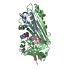
| Structure viewer | Molecule: MolmilJmol/JSmol |
|---|
- Downloads & links
-Download
| PDBx/mmCIF format | 3s2u.cif.gz | 138.3 KB | Display | PDBx/mmCIF format |
|---|---|---|---|---|
| PDB format | pdb3s2u.ent.gz | 108.1 KB | Display | PDB format |
| PDBx/mmJSON format | 3s2u.json.gz | Tree view | PDBx/mmJSON format | |
| Others |  Other downloads Other downloads |
-Validation report
| Arichive directory | https://data.pdbj.org/pub/pdb/validation_reports/s2/3s2uftp://data.pdbj.org/pub/pdb/validation_reports/s2/3s2u | HTTPS FTP |
|---|
-Related structure data
| Similar structure data |
|---|


-Links
 PDBj
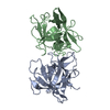
PDBj- Assembly
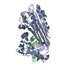
| Deposited unit | 
| ||||||||
|---|---|---|---|---|---|---|---|---|---|
| 1 |
| ||||||||
| Unit cell |
|
-Components
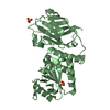
| #1: Protein | Mass: 38919.055 Da / Num. of mol.: 1 Source method: isolated from a genetically manipulated source Source: (gene. exp.) Pseudomonas aeruginosa (bacteria) / Strain: ATCC 15692 / PAO1 / 1C / PRS 101 / LMG 12228 / Gene: murG, PA4412References: UniProt: Q9HW01, undecaprenyldiphospho-muramoylpentapeptide beta-N-acetylglucosaminyltransferase |
|---|---|
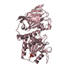
| #2: Chemical | ChemComp-UD1 /   Mass: 607.354 Da / Num. of mol.: 1 / Source method: obtained synthetically / Formula: C17H27N3O17P2 Mass: 607.354 Da / Num. of mol.: 1 / Source method: obtained synthetically / Formula: C17H27N3O17P2 |
| #3: Water | ChemComp-HOH / Water Mass: 18.015 Da / Num. of mol.: 115 / Source method: isolated from a natural source / Formula: H2O Mass: 18.015 Da / Num. of mol.: 115 / Source method: isolated from a natural source / Formula: H2O |
-Experimental details
-Experiment
| Experiment | Method: X-RAY DIFFRACTION / Number of used crystals: 1 |
|---|
- Sample preparation
| Crystal | Density Matthews: 2.23 Å3/Da / Density % sol: 44.87 % |
|---|---|
| Crystal grow | Temperature: 293 K / Method: vapor diffusion / pH: 7 Details: 18% PEG3350, 0.1M HEPES pH 7.0 and 10mM DTT, VAPOR DIFFUSION, temperature 293K |
-Data collection
| Diffraction source | Source: SYNCHROTRON / Site: Diamond  / Beamline: I02 / Beamline: I02 |
|---|---|
| Detector | Type: ADSC QUANTUM 315 / Detector: CCD |
| Radiation | Protocol: SINGLE WAVELENGTH / Monochromatic (M) / Laue (L): M / Scattering type: x-ray |
| Radiation wavelength | Relative weight: 1 |
| Reflection | Resolution: 2.2→20 Å / Num. obs: 15355 / % possible obs: 84.2 % / Observed criterion σ(F): 3 / Observed criterion σ(I): 3 |
- Processing
| Software |
| ||||||||||||||||||||||||||||||||||||||||||||||||||||||||||||||||||||||||||||||||||||||||||||||||||||||||||||||||||
|---|---|---|---|---|---|---|---|---|---|---|---|---|---|---|---|---|---|---|---|---|---|---|---|---|---|---|---|---|---|---|---|---|---|---|---|---|---|---|---|---|---|---|---|---|---|---|---|---|---|---|---|---|---|---|---|---|---|---|---|---|---|---|---|---|---|---|---|---|---|---|---|---|---|---|---|---|---|---|---|---|---|---|---|---|---|---|---|---|---|---|---|---|---|---|---|---|---|---|---|---|---|---|---|---|---|---|---|---|---|---|---|---|---|---|---|
| Refinement | Resolution: 2.23→20 Å / Cor.coef. Fo:Fc: 0.8952 / Cor.coef. Fo:Fc free: 0.8458 / SU R Cruickshank DPI: 0.362 / Cross valid method: THROUGHOUT / σ(F): 0 / SU R Blow DPI: 0.417 / SU Rfree Blow DPI: 0.278 / SU Rfree Cruickshank DPI: 0.271
| ||||||||||||||||||||||||||||||||||||||||||||||||||||||||||||||||||||||||||||||||||||||||||||||||||||||||||||||||||
| Displacement parameters | Biso mean: 43.3 Å2
| ||||||||||||||||||||||||||||||||||||||||||||||||||||||||||||||||||||||||||||||||||||||||||||||||||||||||||||||||||
| Refinement step | Cycle: LAST / Resolution: 2.23→20 Å
| ||||||||||||||||||||||||||||||||||||||||||||||||||||||||||||||||||||||||||||||||||||||||||||||||||||||||||||||||||
| Refine LS restraints |
| ||||||||||||||||||||||||||||||||||||||||||||||||||||||||||||||||||||||||||||||||||||||||||||||||||||||||||||||||||
| LS refinement shell | Resolution: 2.23→2.38 Å / Total num. of bins used: 8
| ||||||||||||||||||||||||||||||||||||||||||||||||||||||||||||||||||||||||||||||||||||||||||||||||||||||||||||||||||
| Refinement TLS params. | Method: refined / Origin x: 10.5945 Å / Origin y: -19.4419 Å / Origin z: -19.1213 Å
| ||||||||||||||||||||||||||||||||||||||||||||||||||||||||||||||||||||||||||||||||||||||||||||||||||||||||||||||||||
| Refinement TLS group | Selection details: { A|* } |
