[English] 日本語
Yorodumi
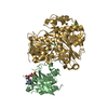
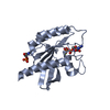
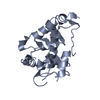
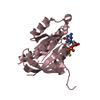
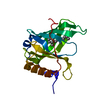
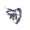
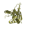
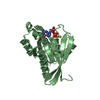
- PDB-3rwo: Crystal structure of YPT32 in complex with GDP -

+
Open data


ID or keywords:

Loading...

-
Basic information

Entry
Database: PDB / ID: 3rwo
TitleCrystal structure of YPT32 in complex with GDP
ComponentsGTP-binding protein YPT32/YPT11
KeywordsPROTEIN TRANSPORT / GTPASES / PROTEIN-GDP COMPLEX / EXOCYTOSIS / GOLGI APPARATUS / GTP-BINDING / LIPOPROTEIN / MEMBRANE / NUCLEOTIDE-BINDING / PRENYLATION / TRANSPORT / Ypt32 / Rab GTPase / GDP / vesicle trafficking / Myo2p / effectors
Function / homology
Function and homology information


Anchoring of the basal body to the plasma membrane / RAB geranylgeranylation / early endosome to Golgi transport / cellular bud neck / exocytosis / vesicle-mediated transport / recycling endosome / autophagy / protein transport / mitochondrial outer membrane ...Anchoring of the basal body to the plasma membrane / RAB geranylgeranylation / early endosome to Golgi transport / cellular bud neck / exocytosis / vesicle-mediated transport / recycling endosome / autophagy / protein transport / mitochondrial outer membrane / endosome / Golgi membrane / GTPase activity / GTP binding / Golgi apparatus / cytosol
Similarity search - Function
small GTPase Rab1 family profile. / Ran (Ras-related nuclear proteins) /TC4 subfamily of small GTPases / Rho (Ras homology) subfamily of Ras-like small GTPases / Ras subfamily of RAS small GTPases / Small GTPase / Ras family / Rab subfamily of small GTPases / Small GTP-binding protein domain / P-loop containing nucleotide triphosphate hydrolases / P-loop containing nucleoside triphosphate hydrolase ...small GTPase Rab1 family profile. / Ran (Ras-related nuclear proteins) /TC4 subfamily of small GTPases / Rho (Ras homology) subfamily of Ras-like small GTPases / Ras subfamily of RAS small GTPases / Small GTPase / Ras family / Rab subfamily of small GTPases / Small GTP-binding protein domain / P-loop containing nucleotide triphosphate hydrolases / P-loop containing nucleoside triphosphate hydrolase / Rossmann fold / 3-Layer(aba) Sandwich / Alpha Beta
Similarity search - Domain/homology
GUANOSINE-5'-DIPHOSPHATE / GTP-binding protein YPT32/YPT11
Similarity search - Component
Biological speciesSaccharomyces cerevisiae (brewer's yeast)
MethodX-RAY DIFFRACTION / SYNCHROTRON / MOLECULAR REPLACEMENT / Resolution: 1.7 Å
AuthorsSultana, A. / Jin, Y. / Dregger, C. / Franklin, E. / Weisman, L.S. / Khan, A.R.
CitationJournal: Febs Lett. / Year: 2011
Title: The activation cycle of Rab GTPase Ypt32 reveals structural determinants of effector recruitment and GDI binding.
Authors: Sultana, A. / Jin, Y. / Dregger, C. / Franklin, E. / Weisman, L.S. / Khan, A.R.
History
DepositionMay 9, 2011Deposition site: RCSB / Processing site: RCSB
Revision 1.0Oct 26, 2011Provider: repository / Type: Initial release
Revision 1.1Nov 9, 2011Group: Database references
Revision 1.2Dec 14, 2011Group: Database references
Revision 1.3Sep 13, 2023Group: Data collection / Database references ...Data collection / Database references / Derived calculations / Refinement description
Category: chem_comp_atom / chem_comp_bond ...chem_comp_atom / chem_comp_bond / database_2 / pdbx_initial_refinement_model / pdbx_struct_conn_angle / struct_conn / struct_ref_seq_dif / struct_site
Item: _database_2.pdbx_DOI / _database_2.pdbx_database_accession ..._database_2.pdbx_DOI / _database_2.pdbx_database_accession / _pdbx_struct_conn_angle.ptnr1_auth_asym_id / _pdbx_struct_conn_angle.ptnr1_auth_comp_id / _pdbx_struct_conn_angle.ptnr1_auth_seq_id / _pdbx_struct_conn_angle.ptnr1_label_asym_id / _pdbx_struct_conn_angle.ptnr1_label_atom_id / _pdbx_struct_conn_angle.ptnr1_label_comp_id / _pdbx_struct_conn_angle.ptnr1_label_seq_id / _pdbx_struct_conn_angle.ptnr2_auth_asym_id / _pdbx_struct_conn_angle.ptnr2_auth_seq_id / _pdbx_struct_conn_angle.ptnr2_label_asym_id / _pdbx_struct_conn_angle.ptnr3_auth_asym_id / _pdbx_struct_conn_angle.ptnr3_auth_comp_id / _pdbx_struct_conn_angle.ptnr3_auth_seq_id / _pdbx_struct_conn_angle.ptnr3_label_asym_id / _pdbx_struct_conn_angle.ptnr3_label_atom_id / _pdbx_struct_conn_angle.ptnr3_label_comp_id / _pdbx_struct_conn_angle.ptnr3_label_seq_id / _pdbx_struct_conn_angle.value / _struct_conn.pdbx_dist_value / _struct_conn.ptnr1_auth_asym_id / _struct_conn.ptnr1_auth_comp_id / _struct_conn.ptnr1_auth_seq_id / _struct_conn.ptnr1_label_asym_id / _struct_conn.ptnr1_label_atom_id / _struct_conn.ptnr1_label_comp_id / _struct_conn.ptnr1_label_seq_id / _struct_conn.ptnr2_auth_asym_id / _struct_conn.ptnr2_auth_comp_id / _struct_conn.ptnr2_auth_seq_id / _struct_conn.ptnr2_label_asym_id / _struct_conn.ptnr2_label_atom_id / _struct_conn.ptnr2_label_comp_id / _struct_ref_seq_dif.details / _struct_site.pdbx_auth_asym_id / _struct_site.pdbx_auth_comp_id / _struct_site.pdbx_auth_seq_id

-
Structure visualization

Structure viewerMolecule:
MolmilJmol/JSmol

Downloads & links

-
Assembly

Deposited unit
B: GTP-binding protein YPT32/YPT11
A: GTP-binding protein YPT32/YPT11
hetero molecules


Theoretical massNumber of molelcules
Total (without water)42,1416
Polymers41,2062
Non-polymers9354
Water8,917495
1
B: GTP-binding protein YPT32/YPT11
hetero molecules


Theoretical massNumber of molelcules
Total (without water)21,0713
Polymers20,6031
Non-polymers4682
Water181
TypeNameSymmetry operationNumber
identity operation1_555x,y,z1
2
A: GTP-binding protein YPT32/YPT11
hetero molecules


Theoretical massNumber of molelcules
Total (without water)21,0713
Polymers20,6031
Non-polymers4682
Water181
TypeNameSymmetry operationNumber
identity operation1_555x,y,z1
3


  • Idetical with deposited unit
  • defined by software
TypeNameSymmetry operationNumber
identity operation1_555x,y,z1
Buried area3120 Å2
ΔGint-47 kcal/mol
Surface area16910 Å2
MethodPISA
Unit cell
Length a, b, c (Å)130.000, 45.200, 73.400
Angle α, β, γ (deg.)90.00, 110.50, 90.00
Int Tables number5
Space group name H-MC121
DetailsAUTHORS STATE THAT THE QUATERNARY STRUCTURE OF YPT32 IN SOLUTION IS A MONOMER. THE DIMER THAT WAS OBSERVED IN THE STRUCTURE IS A RESULT OF CRYSTALLOGRAPHIC PACKING.

-
Components

#1: Protein GTP-binding protein YPT32/YPT11 / Rab GTPase YPT32


Mass: 20603.062 Da / Num. of mol.: 2 / Mutation: Q72L
Source method: isolated from a genetically manipulated source
Source: (gene. exp.) Saccharomyces cerevisiae (brewer's yeast)
Strain: ATCC 204508 / S288c / Gene: YGL210W, YPT11, YPT32 / Production host: Escherichia coli (E. coli) / Strain (production host): BL21 / References: UniProt: P51996
#2: Chemical ChemComp-GDP / GUANOSINE-5'-DIPHOSPHATE / Guanosine diphosphate


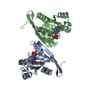
Type: RNA linking / Mass: 443.201 Da / Num. of mol.: 2 / Source method: obtained synthetically / Formula: C10H15N5O11P2 / Comment: GDP, energy-carrying molecule*YM
#3: Chemical ChemComp-MG / MAGNESIUM ION


Mass: 24.305 Da / Num. of mol.: 2 / Source method: obtained synthetically / Formula: Mg
#4: Water ChemComp-HOH / water / Water


Mass: 18.015 Da / Num. of mol.: 495 / Source method: isolated from a natural source / Formula: H2O

-
Experimental details

-
Experiment

ExperimentMethod: X-RAY DIFFRACTION / Number of used crystals: 1

-
Sample preparation

CrystalDensity Matthews: 2.45 Å3/Da / Density % sol: 49.82 %
Crystal growTemperature: 293 K / Method: vapor diffusion, hanging drop / pH: 8.5
Details: PEG 4000, MGCL2, pH 8.5, VAPOR DIFFUSION, HANGING DROP, temperature 293K

-
Data collection

DiffractionMean temperature: 100 K
Diffraction sourceSource: SYNCHROTRON / Site: ESRF / Beamline: BM14 / Wavelength: 0.97849
DetectorType: MARMOSAIC 225 mm CCD / Detector: CCD / Date: Feb 24, 2009
RadiationMonochromator: Si (111) / Protocol: SINGLE WAVELENGTH / Monochromatic (M) / Laue (L): M / Scattering type: x-ray
Radiation wavelengthWavelength: 0.97849 Å / Relative weight: 1
ReflectionResolution: 1.7→68.75 Å / Num. obs: 44104 / % possible obs: 99.4 % / Observed criterion σ(I): -3 / Biso Wilson estimate: 24.66 Å2 / Rmerge(I) obs: 0.052
Reflection shellResolution: 1.7→1.75 Å / Rmerge(I) obs: 0.401 / Mean I/σ(I) obs: 3.9 / % possible all: 98.9

-
Processing

Software
NameVersionClassification
MxCuBEdata collection
MOLREPphasing
REFMAC5.5.0044refinement
XDSdata reduction
XSCALEdata scaling
RefinementMethod to determine structure: MOLECULAR REPLACEMENT
Starting model: PDB ENTRY 3CPJ
Resolution: 1.7→68.75 Å / Cor.coef. Fo:Fc: 0.961 / Cor.coef. Fo:Fc free: 0.943 / SU B: 3.362 / SU ML: 0.059 / Cross valid method: THROUGHOUT / σ(F): 0 / ESU R Free: 0.099 / Stereochemistry target values: MAXIMUM LIKELIHOOD
RfactorNum. reflection% reflectionSelection details
Rfree0.203 2223 5 %RANDOM
Rwork0.167 ---
obs0.169 44104 99.5 %-
Solvent computationIon probe radii: 0.8 Å / Shrinkage radii: 0.8 Å / VDW probe radii: 1.4 Å / Solvent model: MASK
Displacement parametersBiso mean: 20.173 Å2
Baniso -1Baniso -2Baniso -3
1--0.37 Å2-0 Å2-0.08 Å2
2--0.15 Å20 Å2
3---0.17 Å2
Refinement stepCycle: LAST / Resolution: 1.7→68.75 Å
ProteinNucleic acidLigandSolventTotal
Num. atoms2700 0 58 495 3253
Refine LS restraints
Refine-IDTypeDev idealDev ideal targetNumber
X-RAY DIFFRACTIONr_bond_refined_d0.0110.0222907
X-RAY DIFFRACTIONr_angle_refined_deg1.3331.973984
X-RAY DIFFRACTIONr_dihedral_angle_1_deg5.7535380
X-RAY DIFFRACTIONr_dihedral_angle_2_deg37.15925.035143
X-RAY DIFFRACTIONr_dihedral_angle_3_deg14.31615495
X-RAY DIFFRACTIONr_dihedral_angle_4_deg18.0071516
X-RAY DIFFRACTIONr_chiral_restr0.1090.2461
X-RAY DIFFRACTIONr_gen_planes_refined0.0050.022208
X-RAY DIFFRACTIONr_mcbond_it0.7781.51757
X-RAY DIFFRACTIONr_mcangle_it1.41822847
X-RAY DIFFRACTIONr_scbond_it1.94431150
X-RAY DIFFRACTIONr_scangle_it3.0294.51115
LS refinement shellResolution: 1.7→1.74 Å / Total num. of bins used: 20
RfactorNum. reflection% reflection
Rfree0.284 156 -
Rwork0.242 3087 -
obs--98.78 %
Refinement TLS params.

Method: refined / Refine-ID: X-RAY DIFFRACTION

IDL112)L122)L132)L222)L232)L332)S11 (Å °)S12 (Å °)S13 (Å °)S21 (Å °)S22 (Å °)S23 (Å °)S31 (Å °)S32 (Å °)S33 (Å °)T112)T122)T132)T222)T232)T332)Origin x (Å)Origin y (Å)Origin z (Å)
10.3109-0.0817-0.28080.2973-0.1090.67860.02720.00940.00780.0045-0.0376-0.01980.03060.00480.01030.0243-0.0012-0.00810.0106-0.00980.03222.909-7.30345.407
21.8654-1.45321.51121.1672-1.331.9537-0.04870.15610.356-0.0007-0.1744-0.27570.01290.31390.22310.119-0.01890.0190.08480.01580.13069.892-9.74553.775
35.66581.4315-1.16330.8177-0.57480.45220.0368-0.4353-0.26960.178-0.1581-0.1692-0.02940.13360.12130.2305-0.0023-0.02610.1221-0.00680.096211.842-8.9641.586
40.60550.2993-0.34051.3292-0.76780.92890.05590.0557-0.0122-0.0096-0.0675-0.05670.0431-0.00070.01150.03750.0003-0.00350.0209-0.00970.03421.469-6.14844.427
53.0462-0.3546-0.0471.777-1.0431.0183-0.0340.1690.05070.06530.05710.07190.0135-0.2984-0.02320.0288-0.0293-0.00250.1721-0.00790.0805-6.454-4.55143.797
60.473-0.0334-0.2280.5493-0.17421.40150.04890.05920.00920.02060.00920.03570.0153-0.1935-0.05810.01010.0044-0.00960.0358-0.01150.0545-3.028-6.27244.462
70.5708-0.1182-0.09380.6250.10090.82950.04760.0265-0.00560.0226-0.0566-0.02990.0319-0.00880.0090.01020.0028-0.01190.0091-0.00890.03815.075-8.67345.453
Refinement TLS group
IDRefine-IDRefine TLS-IDAuth asym-IDAuth seq-ID
1X-RAY DIFFRACTION1A10 - 35
2X-RAY DIFFRACTION1B10 - 35
3X-RAY DIFFRACTION2A36 - 47
4X-RAY DIFFRACTION3B36 - 47
5X-RAY DIFFRACTION4A48 - 68
6X-RAY DIFFRACTION4B48 - 68
7X-RAY DIFFRACTION5A69 - 78
8X-RAY DIFFRACTION5B69 - 78
9X-RAY DIFFRACTION6A79 - 112
10X-RAY DIFFRACTION6B79 - 112
11X-RAY DIFFRACTION7A121 - 178
12X-RAY DIFFRACTION7B121 - 178

+
About Yorodumi

-
News

-
Feb 9, 2022. New format data for meta-information of EMDB entries

New format data for meta-information of EMDB entries

  • Version 3 of the EMDB header file is now the official format.
  • The previous official version 1.9 will be removed from the archive.

Related info.:EMDB header

External links:wwPDB to switch to version 3 of the EMDB data model

-
Aug 12, 2020. Covid-19 info

Covid-19 info

URL: https://pdbj.org/emnavi/covid19.php

New page: Covid-19 featured information page in EM Navigator.

Related info.:Covid-19 info / Mar 5, 2020. Novel coronavirus structure data

+
Mar 5, 2020. Novel coronavirus structure data

Novel coronavirus structure data

Related info.:Yorodumi Speices / Aug 12, 2020. Covid-19 info

External links:COVID-19 featured content - PDBj / Molecule of the Month (242):Coronavirus Proteases

+
Jan 31, 2019. EMDB accession codes are about to change! (news from PDBe EMDB page)

EMDB accession codes are about to change! (news from PDBe EMDB page)

  • The allocation of 4 digits for EMDB accession codes will soon come to an end. Whilst these codes will remain in use, new EMDB accession codes will include an additional digit and will expand incrementally as the available range of codes is exhausted. The current 4-digit format prefixed with “EMD-” (i.e. EMD-XXXX) will advance to a 5-digit format (i.e. EMD-XXXXX), and so on. It is currently estimated that the 4-digit codes will be depleted around Spring 2019, at which point the 5-digit format will come into force.
  • The EM Navigator/Yorodumi systems omit the EMD- prefix.

Related info.:Q: What is EMD? / ID/Accession-code notation in Yorodumi/EM Navigator

External links:EMDB Accession Codes are Changing Soon! / Contact to PDBj

+
Jul 12, 2017. Major update of PDB

Major update of PDB

  • wwPDB released updated PDB data conforming to the new PDBx/mmCIF dictionary.
  • This is a major update changing the version number from 4 to 5, and with Remediation, in which all the entries are updated.
  • In this update, many items about electron microscopy experimental information are reorganized (e.g. em_software).
  • Now, EM Navigator and Yorodumi are based on the updated data.

External links:wwPDB Remediation / Enriched Model Files Conforming to OneDep Data Standards Now Available in the PDB FTP Archive

-
Yorodumi

Thousand views of thousand structures

  • Yorodumi is a browser for structure data from EMDB, PDB, SASBDB, etc.
  • This page is also the successor to EM Navigator detail page, and also detail information page/front-end page for Omokage search.
  • The word "yorodu" (or yorozu) is an old Japanese word meaning "ten thousand". "mi" (miru) is to see.

Related info.:EMDB / PDB / SASBDB / Comparison of 3 databanks / Yorodumi Search / Aug 31, 2016. New EM Navigator & Yorodumi / Yorodumi Papers / Jmol/JSmol / Function and homology information / Changes in new EM Navigator and Yorodumi

Read more