 Movie
Movie Controller

[English] 日本語
 Yorodumi
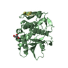
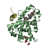
- PDB-3rux: Crystal structure of biotin-protein ligase BirA from Mycobacteriu... -
+ Open data

- Basic information
| Entry | Database: PDB / ID: 3rux | ||||||
|---|---|---|---|---|---|---|---|
| Title | Crystal structure of biotin-protein ligase BirA from Mycobacterium tuberculosis in complex with an acylsulfamide bisubstrate inhibitor | ||||||
 Components Components | BirA bifunctional protein | ||||||
 Keywords Keywords | LIGASE/LIGASE INHIBITOR / biotin-protein ligase / LIGASE-LIGASE INHIBITOR complex | ||||||
| Function / homology |  Function and homology information Function and homology informationbiotin-protein ligase activity / biotin-[biotin carboxyl-carrier protein] ligase / biotin-[acetyl-CoA-carboxylase] ligase activity /  small molecule binding / post-translational protein modification small molecule binding / post-translational protein modificationSimilarity search - Function | ||||||
| Biological species |   Mycobacterium tuberculosis (bacteria) Mycobacterium tuberculosis (bacteria) | ||||||
| Method | X-RAY DIFFRACTION / SYNCHROTRON / Resolution: 1.7 Å | ||||||
 Authors Authors | Geders, T.W. / Finzel, B.C. | ||||||
 Citation Citation | Journal: Chem.Biol. / Year: 2011 Title: Bisubstrate Adenylation Inhibitors of Biotin Protein Ligase from Mycobacterium tuberculosis. Authors: Duckworth, B.P. / Geders, T.W. / Tiwari, D. / Boshoff, H.I. / Sibbald, P.A. / Barry, C.E. / Schnappinger, D. / Finzel, B.C. / Aldrich, C.C. | ||||||
| History |
|
- Structure visualization
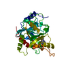
| Structure viewer | Molecule: MolmilJmol/JSmol |
|---|
- Downloads & links
-Download
| PDBx/mmCIF format | 3rux.cif.gz | 130.6 KB | Display | PDBx/mmCIF format |
|---|---|---|---|---|
| PDB format | pdb3rux.ent.gz | 99.7 KB | Display | PDB format |
| PDBx/mmJSON format | 3rux.json.gz | Tree view | PDBx/mmJSON format | |
| Others |  Other downloads Other downloads |
-Validation report
| Arichive directory | https://data.pdbj.org/pub/pdb/validation_reports/ru/3ruxftp://data.pdbj.org/pub/pdb/validation_reports/ru/3rux | HTTPS FTP |
|---|
-Related structure data
| Related structure data | |
|---|---|
| Similar structure data |


-Links
 PDBj
PDBj
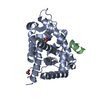
- Assembly
| Deposited unit | 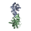
| ||||||||
|---|---|---|---|---|---|---|---|---|---|
| 1 | 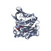
| ||||||||
| 2 | 
| ||||||||
| Unit cell |
|
-Components
| #1: Protein | Mass: 28534.428 Da / Num. of mol.: 2 Source method: isolated from a genetically manipulated source Source: (gene. exp.) Mycobacterium tuberculosis (bacteria) / Strain: H37Rv / Gene: birA, MT3379, Rv3279c / Plasmid: pET-28b / Production host: Escherichia coli (E. coli) / Strain (production host): MACH IReferences: UniProt: P96884, biotin-[biotin carboxyl-carrier protein] ligase #2: Chemical |   Mass: 571.630 Da / Num. of mol.: 2 / Source method: obtained synthetically / Formula: C20H29N9O7S2 Mass: 571.630 Da / Num. of mol.: 2 / Source method: obtained synthetically / Formula: C20H29N9O7S2#3: Water | ChemComp-HOH / | Water Mass: 18.015 Da / Num. of mol.: 653 / Source method: isolated from a natural source / Formula: H2O Mass: 18.015 Da / Num. of mol.: 653 / Source method: isolated from a natural source / Formula: H2O |
|---|
-Experimental details
-Experiment
| Experiment | Method: X-RAY DIFFRACTION / Number of used crystals: 1 |
|---|
- Sample preparation
| Crystal | Density Matthews: 2.09 Å3/Da / Density % sol: 41.16 % |
|---|---|
| Crystal grow | Temperature: 293.15 K / Method: vapor diffusion, hanging drop / pH: 8.5 Details: 20% MPEG 2000, 50 mM trimethylamine N-oxide, 100 mM Tris, 20% PEG 400 as cryoprotectant, VAPOR DIFFUSION, HANGING DROP, temperature 293.15K, pH 8.5 |
-Data collection
| Diffraction | Mean temperature: 100 K |
|---|---|
| Diffraction source | Source: SYNCHROTRON / Site: ALS  / Beamline: 4.2.2 / Wavelength: 1 Å / Beamline: 4.2.2 / Wavelength: 1 Å |
| Detector | Type: NOIR-1 / Detector: CCD / Date: Dec 16, 2009 |
| Radiation | Monochromator: Rosenbaum-Rock monochromator Si 111 / Protocol: SINGLE WAVELENGTH / Monochromatic (M) / Laue (L): M / Scattering type: x-ray |
| Radiation wavelength | Wavelength: 1 Å / Relative weight: 1 |
| Reflection | Resolution: 1.7→43.399 Å / Num. all: 56877 / Num. obs: 55584 / % possible obs: 97.7 % / Observed criterion σ(F): 1 / Observed criterion σ(I): 1 / Redundancy: 6.8 % / Biso Wilson estimate: 21.9 Å2 / Rmerge(I) obs: 0.06 / Net I/σ(I): 15.6 |
- Processing
| Software |
| |||||||||||||||||||||||||||||||||||||||||||||||||||||||||||||||||||||||||||||
|---|---|---|---|---|---|---|---|---|---|---|---|---|---|---|---|---|---|---|---|---|---|---|---|---|---|---|---|---|---|---|---|---|---|---|---|---|---|---|---|---|---|---|---|---|---|---|---|---|---|---|---|---|---|---|---|---|---|---|---|---|---|---|---|---|---|---|---|---|---|---|---|---|---|---|---|---|---|---|
| Refinement | Resolution: 1.7→43.399 Å / Occupancy max: 1 / Occupancy min: 0 / FOM work R set: 0.83 / SU ML: 0.43 / σ(F): 1.34 / Phase error: 23.38 / Stereochemistry target values: ML
| |||||||||||||||||||||||||||||||||||||||||||||||||||||||||||||||||||||||||||||
| Solvent computation | Shrinkage radii: 0.9 Å / VDW probe radii: 1.11 Å / Solvent model: FLAT BULK SOLVENT MODEL / Bsol: 36.325 Å2 / ksol: 0.31 e/Å3 | |||||||||||||||||||||||||||||||||||||||||||||||||||||||||||||||||||||||||||||
| Displacement parameters | Biso max: 68.99 Å2 / Biso mean: 23.2336 Å2 / Biso min: 8.43 Å2
| |||||||||||||||||||||||||||||||||||||||||||||||||||||||||||||||||||||||||||||
| Refinement step | Cycle: LAST / Resolution: 1.7→43.399 Å
| |||||||||||||||||||||||||||||||||||||||||||||||||||||||||||||||||||||||||||||
| Refine LS restraints |
| |||||||||||||||||||||||||||||||||||||||||||||||||||||||||||||||||||||||||||||
| LS refinement shell | Refine-ID: X-RAY DIFFRACTION / Total num. of bins used: 10
|
