 Movie
Movie Controller


+ Open data

- Basic information
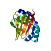
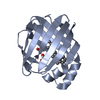
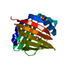
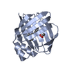
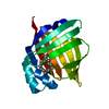
| Entry | Database: PDB / ID: 3rsw | ||||||
|---|---|---|---|---|---|---|---|
| Title | Crystal Structure of Heart Fatty Acid Binding Protein (FABP3) | ||||||
 Components Components | Fatty acid-binding protein, heart | ||||||
 Keywords Keywords |  CHAPERONE / Lipid carrier / molecular chaperone / Heart Fatty Acid Binding Protein / Type 2 diabetes / atherosclerosis CHAPERONE / Lipid carrier / molecular chaperone / Heart Fatty Acid Binding Protein / Type 2 diabetes / atherosclerosis | ||||||
| Function / homology |  Function and homology information Function and homology informationpositive regulation of long-chain fatty acid import into cell / regulation of phosphatidylcholine biosynthetic process / regulation of fatty acid oxidation / positive regulation of phospholipid biosynthetic process / intracellular lipid transport / oleic acid binding / phospholipid homeostasis / long-chain fatty acid binding / Triglyceride catabolism / long-chain fatty acid transport ...positive regulation of long-chain fatty acid import into cell / regulation of phosphatidylcholine biosynthetic process / regulation of fatty acid oxidation / positive regulation of phospholipid biosynthetic process / intracellular lipid transport / oleic acid binding / phospholipid homeostasis / long-chain fatty acid binding / Triglyceride catabolism / long-chain fatty acid transport / brown fat cell differentiation / cytoskeletal protein binding / cholesterol homeostasis / negative regulation of cell population proliferation / extracellular space / extracellular exosome / nucleus / cytosolSimilarity search - Function | ||||||
| Biological species |  Homo sapiens (human) Homo sapiens (human) | ||||||
| Method | X-RAY DIFFRACTION / SYNCHROTRON / MOLECULAR REPLACEMENT / Resolution: 2.599 Å | ||||||
 Authors Authors | Carney, D.F. / Shankaran, B. / Zwart, P.H. / Stebbins, J.W. / Prasad, G.S. | ||||||
 Citation Citation | Journal: To be Published Title: Crystal Structure of Heart Fatty Acid Binding Protein (FABP3) Authors: Carney, D.F. / Shankaran, B. / Zwart, P.H. / Stebbins, J.W. / Prasad, G.S. | ||||||
| History |
|
- Structure visualization
| Structure viewer | Molecule: MolmilJmol/JSmol |
|---|
- Downloads & links
-Download
| PDBx/mmCIF format | 3rsw.cif.gz | 116.8 KB | Display | PDBx/mmCIF format |
|---|---|---|---|---|
| PDB format | pdb3rsw.ent.gz | 91.5 KB | Display | PDB format |
| PDBx/mmJSON format | 3rsw.json.gz | Tree view | PDBx/mmJSON format | |
| Others |  Other downloads Other downloads |
-Validation report
| Arichive directory | https://data.pdbj.org/pub/pdb/validation_reports/rs/3rswftp://data.pdbj.org/pub/pdb/validation_reports/rs/3rsw | HTTPS FTP |
|---|
-Related structure data
| Related structure data | |
|---|---|
| Similar structure data |


-Links
 PDBj
PDBj


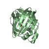
- Assembly
| Deposited unit | 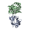
| ||||||||
|---|---|---|---|---|---|---|---|---|---|
| 1 | 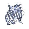
| ||||||||
| 2 | 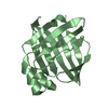
| ||||||||
| Unit cell |
|
-Components
| #1: Protein | Mass: 17651.123 Da / Num. of mol.: 2 Source method: isolated from a genetically manipulated source Source: (gene. exp.) Homo sapiens (human) / Gene: FABP3, FABP11, MDGI / Plasmid: pET15b / Production host:  Escherichia coli (E. coli) / Strain (production host): BL21 / References: UniProt: P05413 Escherichia coli (E. coli) / Strain (production host): BL21 / References: UniProt: P05413#2: Water | ChemComp-HOH / | Water Mass: 18.015 Da / Num. of mol.: 37 / Source method: isolated from a natural source / Formula: H2O Mass: 18.015 Da / Num. of mol.: 37 / Source method: isolated from a natural source / Formula: H2O |
|---|
-Experimental details
-Experiment
| Experiment | Method: X-RAY DIFFRACTION |
|---|
- Sample preparation
| Crystal | Density Matthews: 2.16 Å3/Da / Density % sol: 42.98 % |
|---|---|
| Crystal grow | Temperature: 298 K / Method: vapor diffusion, sitting drop / pH: 3.2 Details: 0.1M Citric Acid, pH 3.2 and 32% PEG400, VAPOR DIFFUSION, SITTING DROP, temperature 298K |
-Data collection
| Diffraction | Mean temperature: 80 K | |||||||||||||||
|---|---|---|---|---|---|---|---|---|---|---|---|---|---|---|---|---|
| Diffraction source | Source: SYNCHROTRON / Site: ALS  / Beamline: 5.0.1 / Wavelength: 0.9774 Å / Beamline: 5.0.1 / Wavelength: 0.9774 Å | |||||||||||||||
| Detector | Type: ADSC QUANTUM 315 / Detector: CCD / Date: Apr 28, 2011 | |||||||||||||||
| Radiation | Protocol: SINGLE WAVELENGTH / Monochromatic (M) / Laue (L): M / Scattering type: x-ray | |||||||||||||||
| Radiation wavelength | Wavelength: 0.9774 Å / Relative weight: 1 | |||||||||||||||
| Reflection twin |
| |||||||||||||||
| Reflection | Resolution: 2.599→84.4 Å / Num. all: 9216 / Num. obs: 9216 / % possible obs: 99.9 % / Observed criterion σ(F): 1.5 / Observed criterion σ(I): 1.5 / Redundancy: 11.4 % / Biso Wilson estimate: 62.5 Å2 / Rmerge(I) obs: 0.167 / Rsym value: 0.167 / Net I/σ(I): 11.8 |
- Processing
| Software |
| |||||||||||||||||||||||||||||||||||||||||||||||||||||||||||||||||||||||||||
|---|---|---|---|---|---|---|---|---|---|---|---|---|---|---|---|---|---|---|---|---|---|---|---|---|---|---|---|---|---|---|---|---|---|---|---|---|---|---|---|---|---|---|---|---|---|---|---|---|---|---|---|---|---|---|---|---|---|---|---|---|---|---|---|---|---|---|---|---|---|---|---|---|---|---|---|---|
| Refinement | Method to determine structure: MOLECULAR REPLACEMENT / Resolution: 2.599→42.48 Å / Cor.coef. Fo:Fc: 0.923 / Cor.coef. Fo:Fc free: 0.896 / SU B: 20.78 / SU ML: 0.208 / Cross valid method: THROUGHOUT / ESU R: 0.341 / ESU R Free: 0.075 / Stereochemistry target values: MAXIMUM LIKELIHOOD / Details: HYDROGENS HAVE BEEN ADDED IN THE RIDING POSITIONS
| |||||||||||||||||||||||||||||||||||||||||||||||||||||||||||||||||||||||||||
| Solvent computation | Ion probe radii: 0.8 Å / Shrinkage radii: 0.8 Å / VDW probe radii: 1.4 Å / Solvent model: MASK | |||||||||||||||||||||||||||||||||||||||||||||||||||||||||||||||||||||||||||
| Displacement parameters | Biso mean: 50.761 Å2
| |||||||||||||||||||||||||||||||||||||||||||||||||||||||||||||||||||||||||||
| Refinement step | Cycle: LAST / Resolution: 2.599→42.48 Å
| |||||||||||||||||||||||||||||||||||||||||||||||||||||||||||||||||||||||||||
| Refine LS restraints |
| |||||||||||||||||||||||||||||||||||||||||||||||||||||||||||||||||||||||||||
| LS refinement shell | Resolution: 2.599→2.666 Å / Total num. of bins used: 20
| |||||||||||||||||||||||||||||||||||||||||||||||||||||||||||||||||||||||||||
| Refinement TLS params. | Method: refined / Refine-ID: X-RAY DIFFRACTION
| |||||||||||||||||||||||||||||||||||||||||||||||||||||||||||||||||||||||||||
| Refinement TLS group |
|
