 Movie
Movie Controller

[English] 日本語
 Yorodumi
- PDB-3rpx: Crystal structure of complement component 1, q subcomponent bindi... -
+ Open data

- Basic information
| Entry | Database: PDB / ID: 3rpx | ||||||
|---|---|---|---|---|---|---|---|
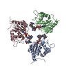
| Title | Crystal structure of complement component 1, q subcomponent binding protein, C1QBP | ||||||
 Components Components | Complement component 1 Q subcomponent-binding protein | ||||||
 Keywords Keywords |  PROTEIN BINDING / structural genomics / structural genomics consortium / mitochondrion matrix / complement system / protein-binding / SGC PROTEIN BINDING / structural genomics / structural genomics consortium / mitochondrion matrix / complement system / protein-binding / SGC | ||||||
| Function / homology |  Function and homology informationadrenergic receptor binding / Apoptotic factor-mediated response / Defective Intrinsic Pathway for Apoptosis Due to p14ARF Loss of Function / negative regulation of MDA-5 signaling pathway / kininogen binding / negative regulation of RIG-I signaling pathway / negative regulation of defense response to virus / positive regulation of dendritic cell chemotaxis / hyaluronic acid binding / complement component C1q complex binding ...adrenergic receptor binding / Apoptotic factor-mediated response / Defective Intrinsic Pathway for Apoptosis Due to p14ARF Loss of Function / negative regulation of MDA-5 signaling pathway / kininogen binding / negative regulation of RIG-I signaling pathway / negative regulation of defense response to virus / positive regulation of dendritic cell chemotaxis / hyaluronic acid binding / complement component C1q complex binding / positive regulation of trophoblast cell migration / mitochondrial ribosome binding / regulation of complement activation / positive regulation of mitochondrial translation / negative regulation of interleukin-12 production / positive regulation of neutrophil chemotaxis / enzyme inhibitor activity / RHOC GTPase cycle / negative regulation of mRNA splicing, via spliceosome / transcription factor binding / negative regulation of type II interferon production / positive regulation of cell adhesion / RHOA GTPase cycle / negative regulation of double-strand break repair via homologous recombination / positive regulation of substrate adhesion-dependent cell spreading / Intrinsic Pathway of Fibrin Clot Formation / complement activation, classical pathway / RNA splicing / phosphatidylinositol 3-kinase/protein kinase B signal transduction / cytosolic ribosome assembly / mRNA processing / transcription corepressor activity / positive regulation of phosphatidylinositol 3-kinase/protein kinase B signal transduction / mitochondrial matrix / immune response / positive regulation of apoptotic process / innate immune response / mRNA binding / apoptotic process / nucleolus / negative regulation of transcription by RNA polymerase II / cell surface / mitochondrion / extracellular region / membrane / nucleus / plasma membrane / cytosol / cytoplasm Function and homology informationadrenergic receptor binding / Apoptotic factor-mediated response / Defective Intrinsic Pathway for Apoptosis Due to p14ARF Loss of Function / negative regulation of MDA-5 signaling pathway / kininogen binding / negative regulation of RIG-I signaling pathway / negative regulation of defense response to virus / positive regulation of dendritic cell chemotaxis / hyaluronic acid binding / complement component C1q complex binding ...adrenergic receptor binding / Apoptotic factor-mediated response / Defective Intrinsic Pathway for Apoptosis Due to p14ARF Loss of Function / negative regulation of MDA-5 signaling pathway / kininogen binding / negative regulation of RIG-I signaling pathway / negative regulation of defense response to virus / positive regulation of dendritic cell chemotaxis / hyaluronic acid binding / complement component C1q complex binding / positive regulation of trophoblast cell migration / mitochondrial ribosome binding / regulation of complement activation / positive regulation of mitochondrial translation / negative regulation of interleukin-12 production / positive regulation of neutrophil chemotaxis / enzyme inhibitor activity / RHOC GTPase cycle / negative regulation of mRNA splicing, via spliceosome / transcription factor binding / negative regulation of type II interferon production / positive regulation of cell adhesion / RHOA GTPase cycle / negative regulation of double-strand break repair via homologous recombination / positive regulation of substrate adhesion-dependent cell spreading / Intrinsic Pathway of Fibrin Clot Formation / complement activation, classical pathway / RNA splicing / phosphatidylinositol 3-kinase/protein kinase B signal transduction / cytosolic ribosome assembly / mRNA processing / transcription corepressor activity / positive regulation of phosphatidylinositol 3-kinase/protein kinase B signal transduction / mitochondrial matrix / immune response / positive regulation of apoptotic process / innate immune response / mRNA binding / apoptotic process / nucleolus / negative regulation of transcription by RNA polymerase II / cell surface / mitochondrion / extracellular region / membrane / nucleus / plasma membrane / cytosol / cytoplasmSimilarity search - Function | ||||||
| Biological species |  Homo sapiens (human) Homo sapiens (human) | ||||||
| Method | X-RAY DIFFRACTION / SYNCHROTRON / MOLECULAR REPLACEMENT / molecular replacement / Resolution: 2.65 Å | ||||||
 Authors Authors | Tempel, W. / Tong, Y. / Crombet, L. / Shen, Y. / Guan, X. / Nedyalkova, L. / Walker, J.R. / Arrowsmith, C.H. / Edwards, A.M. / Bountra, C. ...Tempel, W. / Tong, Y. / Crombet, L. / Shen, Y. / Guan, X. / Nedyalkova, L. / Walker, J.R. / Arrowsmith, C.H. / Edwards, A.M. / Bountra, C. / Weigelt, J. / Park, H. / Structural Genomics Consortium (SGC) | ||||||
 Citation Citation | Journal: to be published Title: Crystal structure of complement component 1, q subcomponent binding protein, C1QBP Authors: Tempel, W. / Tong, Y. / Crombet, L. / Shen, Y. / Guan, X. / Nedyalkova, L. / Walker, J.R. / Arrowsmith, C.H. / Edwards, A.M. / Bountra, C. / Weigelt, J. / Park, H. | ||||||
| History |
|
- Structure visualization
| Structure viewer | Molecule: MolmilJmol/JSmol |
|---|
- Downloads & links
-Download
| PDBx/mmCIF format | 3rpx.cif.gz | 179.7 KB | Display | PDBx/mmCIF format |
|---|---|---|---|---|
| PDB format | pdb3rpx.ent.gz | 142.4 KB | Display | PDB format |
| PDBx/mmJSON format | 3rpx.json.gz | Tree view | PDBx/mmJSON format | |
| Others |  Other downloads Other downloads |
-Validation report
| Arichive directory | https://data.pdbj.org/pub/pdb/validation_reports/rp/3rpxftp://data.pdbj.org/pub/pdb/validation_reports/rp/3rpx | HTTPS FTP |
|---|
-Related structure data
| Related structure data | 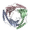 1p32S S: Starting model for refinement |
|---|---|
| Similar structure data |


-Links
 PDBj
PDBj


- Assembly
| Deposited unit | 
| ||||||||
|---|---|---|---|---|---|---|---|---|---|
| 1 |
| ||||||||
| Unit cell |
|
-Components
| #1: Protein | Mass: 22128.186 Da / Num. of mol.: 3 / Fragment: UNP residues 96-282 Source method: isolated from a genetically manipulated source Source: (gene. exp.) Homo sapiens (human) / Gene: C1QBP, GC1QBP, HABP1, SF2P32 / Plasmid: pNIC-CH / Production host:  Escherichia coli (E. coli) / Strain (production host): BL21(DE3)-V2R-pRARE2 / References: UniProt: Q07021 Escherichia coli (E. coli) / Strain (production host): BL21(DE3)-V2R-pRARE2 / References: UniProt: Q07021#2: Chemical | ChemComp-UNX /   Num. of mol.: 19 / Source method: obtained synthetically Num. of mol.: 19 / Source method: obtained syntheticallySequence details | THE ORIGINAL EXPRESSION CONSTRUCT ENCOMPASSED RESIDUES 3-282 OF UNIPROT SEQUENCE Q07021. WE SUSPECT ...THE ORIGINAL EXPRESSION | |
|---|
-Experimental details
-Experiment
| Experiment | Method: X-RAY DIFFRACTION / Number of used crystals: 1 |
|---|
- Sample preparation
| Crystal | Density Matthews: 3.7 Å3/Da / Density % sol: 66.72 % |
|---|---|
| Crystal grow | Temperature: 293 K / Method: vapor diffusion, sitting drop / pH: 7.5 Details: 25% peg1500, 0.2 M sodium chloride, 0.1 M HEPES, 5% glycerol, pH 7.5, VAPOR DIFFUSION, SITTING DROP, temperature 293K |
-Data collection
| Diffraction | Mean temperature: 100 K | |||||||||||||||||||||||||||||||||||||||||||||||||||||||||||||||||||||||||||||||||||||||||||||||||||||||||||||||||||||||||||||||||||||||||||||||||||
|---|---|---|---|---|---|---|---|---|---|---|---|---|---|---|---|---|---|---|---|---|---|---|---|---|---|---|---|---|---|---|---|---|---|---|---|---|---|---|---|---|---|---|---|---|---|---|---|---|---|---|---|---|---|---|---|---|---|---|---|---|---|---|---|---|---|---|---|---|---|---|---|---|---|---|---|---|---|---|---|---|---|---|---|---|---|---|---|---|---|---|---|---|---|---|---|---|---|---|---|---|---|---|---|---|---|---|---|---|---|---|---|---|---|---|---|---|---|---|---|---|---|---|---|---|---|---|---|---|---|---|---|---|---|---|---|---|---|---|---|---|---|---|---|---|---|---|---|---|
| Diffraction source | Source: SYNCHROTRON / Site: CLSI  / Beamline: 08ID-1 / Wavelength: 0.9792 Å / Beamline: 08ID-1 / Wavelength: 0.9792 Å | |||||||||||||||||||||||||||||||||||||||||||||||||||||||||||||||||||||||||||||||||||||||||||||||||||||||||||||||||||||||||||||||||||||||||||||||||||
| Detector | Type: MARMOSAIC 225 mm CCD / Detector: CCD / Date: Sep 12, 2009 | |||||||||||||||||||||||||||||||||||||||||||||||||||||||||||||||||||||||||||||||||||||||||||||||||||||||||||||||||||||||||||||||||||||||||||||||||||
| Radiation | Protocol: SINGLE WAVELENGTH / Monochromatic (M) / Laue (L): M / Scattering type: x-ray | |||||||||||||||||||||||||||||||||||||||||||||||||||||||||||||||||||||||||||||||||||||||||||||||||||||||||||||||||||||||||||||||||||||||||||||||||||
| Radiation wavelength | Wavelength: 0.9792 Å / Relative weight: 1 | |||||||||||||||||||||||||||||||||||||||||||||||||||||||||||||||||||||||||||||||||||||||||||||||||||||||||||||||||||||||||||||||||||||||||||||||||||
| Reflection | Resolution: 2.65→40 Å / Num. obs: 28824 / % possible obs: 99.6 % / Observed criterion σ(I): -3 / Biso Wilson estimate: 67.228 Å2 / Rmerge(I) obs: 0.074 / Net I/σ(I): 18.46 | |||||||||||||||||||||||||||||||||||||||||||||||||||||||||||||||||||||||||||||||||||||||||||||||||||||||||||||||||||||||||||||||||||||||||||||||||||
| Reflection shell |
|
-Phasing
| Phasing | Method: molecular replacement |
|---|
- Processing
| Software |
| ||||||||||||||||||||||||||||||||||||||||||||||||||||||||||||||||||||||||||||||||||||||||||||||||||||||||||||
|---|---|---|---|---|---|---|---|---|---|---|---|---|---|---|---|---|---|---|---|---|---|---|---|---|---|---|---|---|---|---|---|---|---|---|---|---|---|---|---|---|---|---|---|---|---|---|---|---|---|---|---|---|---|---|---|---|---|---|---|---|---|---|---|---|---|---|---|---|---|---|---|---|---|---|---|---|---|---|---|---|---|---|---|---|---|---|---|---|---|---|---|---|---|---|---|---|---|---|---|---|---|---|---|---|---|---|---|---|---|
| Refinement | Method to determine structure: MOLECULAR REPLACEMENT Starting model: pdb entry 1p32 Resolution: 2.65→19.7 Å / Cor.coef. Fo:Fc: 0.94 / Cor.coef. Fo:Fc free: 0.9363 / Occupancy max: 1 / Occupancy min: 0.5 / Cross valid method: THROUGHOUT / σ(F): 0 Details: refmac, phenix, coot and the molprobity server were also used during refinement. UNK atoms were refined as water rather than omitted in order to better approximate the properties of these unknown entities.
| ||||||||||||||||||||||||||||||||||||||||||||||||||||||||||||||||||||||||||||||||||||||||||||||||||||||||||||
| Displacement parameters | Biso max: 164.1 Å2 / Biso mean: 82.1977 Å2 / Biso min: 27.64 Å2
| ||||||||||||||||||||||||||||||||||||||||||||||||||||||||||||||||||||||||||||||||||||||||||||||||||||||||||||
| Refine analyze | Luzzati coordinate error obs: 0.442 Å | ||||||||||||||||||||||||||||||||||||||||||||||||||||||||||||||||||||||||||||||||||||||||||||||||||||||||||||
| Refinement step | Cycle: LAST / Resolution: 2.65→19.7 Å
| ||||||||||||||||||||||||||||||||||||||||||||||||||||||||||||||||||||||||||||||||||||||||||||||||||||||||||||
| Refine LS restraints |
| ||||||||||||||||||||||||||||||||||||||||||||||||||||||||||||||||||||||||||||||||||||||||||||||||||||||||||||
| LS refinement shell | Resolution: 2.65→2.75 Å / Total num. of bins used: 14
| ||||||||||||||||||||||||||||||||||||||||||||||||||||||||||||||||||||||||||||||||||||||||||||||||||||||||||||
| Refinement TLS params. | Method: refined / Refine-ID: X-RAY DIFFRACTION
| ||||||||||||||||||||||||||||||||||||||||||||||||||||||||||||||||||||||||||||||||||||||||||||||||||||||||||||
| Refinement TLS group |
|
