 Movie
Movie Controller

[English] 日本語
 Yorodumi
- PDB-1p32: CRYSTAL STRUCTURE OF HUMAN P32, A DOUGHNUT-SHAPED ACIDIC MITOCHON... -
+ Open data

- Basic information
| Entry | Database: PDB / ID: 1p32 | ||||||
|---|---|---|---|---|---|---|---|
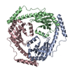
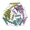
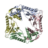
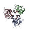
| Title | CRYSTAL STRUCTURE OF HUMAN P32, A DOUGHNUT-SHAPED ACIDIC MITOCHONDRIAL MATRIX PROTEIN | ||||||
 Components Components | MITOCHONDRIAL MATRIX PROTEIN, SF2P32 | ||||||
 Keywords Keywords | MITOCHONDRIAL MATRIX PROTEIN | ||||||
| Function / homology |  Function and homology informationadrenergic receptor binding / Apoptotic factor-mediated response / Defective Intrinsic Pathway for Apoptosis Due to p14ARF Loss of Function / negative regulation of MDA-5 signaling pathway / kininogen binding / negative regulation of RIG-I signaling pathway / : / negative regulation of defense response to virus / positive regulation of dendritic cell chemotaxis / hyaluronic acid binding ...adrenergic receptor binding / Apoptotic factor-mediated response / Defective Intrinsic Pathway for Apoptosis Due to p14ARF Loss of Function / negative regulation of MDA-5 signaling pathway / kininogen binding / negative regulation of RIG-I signaling pathway / : / negative regulation of defense response to virus / positive regulation of dendritic cell chemotaxis / hyaluronic acid binding / complement component C1q complex binding / translation activator activity / positive regulation of trophoblast cell migration / mitochondrial ribosome binding / blood coagulation, intrinsic pathway / regulation of complement activation / positive regulation of mitochondrial translation / negative regulation of interleukin-12 production / positive regulation of neutrophil chemotaxis / enzyme inhibitor activity / presynaptic active zone / RHOC GTPase cycle / negative regulation of mRNA splicing, via spliceosome / transcription factor binding / negative regulation of type II interferon production / positive regulation of cell adhesion / RHOA GTPase cycle / GABA-ergic synapse / negative regulation of double-strand break repair via homologous recombination / positive regulation of substrate adhesion-dependent cell spreading / Intrinsic Pathway of Fibrin Clot Formation / viral process / intrinsic apoptotic signaling pathway / complement activation, classical pathway / RNA splicing / phosphatidylinositol 3-kinase/protein kinase B signal transduction / cytosolic ribosome assembly / protein kinase C binding / mRNA processing / transcription corepressor activity / positive regulation of phosphatidylinositol 3-kinase/protein kinase B signal transduction / mitochondrial matrix / immune response / positive regulation of apoptotic process / innate immune response / mRNA binding / glutamatergic synapse / apoptotic process / nucleolus / negative regulation of transcription by RNA polymerase II / cell surface / mitochondrion / extracellular space / extracellular region / membrane / nucleus / plasma membrane / cytosol / cytoplasm Function and homology informationadrenergic receptor binding / Apoptotic factor-mediated response / Defective Intrinsic Pathway for Apoptosis Due to p14ARF Loss of Function / negative regulation of MDA-5 signaling pathway / kininogen binding / negative regulation of RIG-I signaling pathway / : / negative regulation of defense response to virus / positive regulation of dendritic cell chemotaxis / hyaluronic acid binding ...adrenergic receptor binding / Apoptotic factor-mediated response / Defective Intrinsic Pathway for Apoptosis Due to p14ARF Loss of Function / negative regulation of MDA-5 signaling pathway / kininogen binding / negative regulation of RIG-I signaling pathway / : / negative regulation of defense response to virus / positive regulation of dendritic cell chemotaxis / hyaluronic acid binding / complement component C1q complex binding / translation activator activity / positive regulation of trophoblast cell migration / mitochondrial ribosome binding / blood coagulation, intrinsic pathway / regulation of complement activation / positive regulation of mitochondrial translation / negative regulation of interleukin-12 production / positive regulation of neutrophil chemotaxis / enzyme inhibitor activity / presynaptic active zone / RHOC GTPase cycle / negative regulation of mRNA splicing, via spliceosome / transcription factor binding / negative regulation of type II interferon production / positive regulation of cell adhesion / RHOA GTPase cycle / GABA-ergic synapse / negative regulation of double-strand break repair via homologous recombination / positive regulation of substrate adhesion-dependent cell spreading / Intrinsic Pathway of Fibrin Clot Formation / viral process / intrinsic apoptotic signaling pathway / complement activation, classical pathway / RNA splicing / phosphatidylinositol 3-kinase/protein kinase B signal transduction / cytosolic ribosome assembly / protein kinase C binding / mRNA processing / transcription corepressor activity / positive regulation of phosphatidylinositol 3-kinase/protein kinase B signal transduction / mitochondrial matrix / immune response / positive regulation of apoptotic process / innate immune response / mRNA binding / glutamatergic synapse / apoptotic process / nucleolus / negative regulation of transcription by RNA polymerase II / cell surface / mitochondrion / extracellular space / extracellular region / membrane / nucleus / plasma membrane / cytosol / cytoplasmSimilarity search - Function | ||||||
| Biological species |  Homo sapiens (human) Homo sapiens (human) | ||||||
| Method | X-RAY DIFFRACTION / SYNCHROTRON / MAD / Resolution: 2.25 Å | ||||||
 Authors Authors | Jiang, J. / Zhang, Y. / Krainer, A.R. / Xu, R.-M. | ||||||
 Citation Citation | Journal: Proc.Natl.Acad.Sci.USA / Year: 1999 Title: Crystal structure of human p32, a doughnut-shaped acidic mitochondrial matrix protein. Authors: Jiang, J. / Zhang, Y. / Krainer, A.R. / Xu, R.M. | ||||||
| History |
|
- Structure visualization
| Structure viewer | Molecule: MolmilJmol/JSmol |
|---|
- Downloads & links
-Download
| PDBx/mmCIF format | 1p32.cif.gz | 125.1 KB | Display | PDBx/mmCIF format |
|---|---|---|---|---|
| PDB format | pdb1p32.ent.gz | 97.6 KB | Display | PDB format |
| PDBx/mmJSON format | 1p32.json.gz | Tree view | PDBx/mmJSON format | |
| Others |  Other downloads Other downloads |
-Validation report
| Arichive directory | https://data.pdbj.org/pub/pdb/validation_reports/p3/1p32ftp://data.pdbj.org/pub/pdb/validation_reports/p3/1p32 | HTTPS FTP |
|---|
-Related structure data
| Similar structure data |
|---|


-Links
 PDBj
PDBj


- Assembly
| Deposited unit | 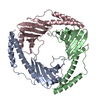
| ||||||||||||||||
|---|---|---|---|---|---|---|---|---|---|---|---|---|---|---|---|---|---|
| 1 |
| ||||||||||||||||
| Unit cell |
| ||||||||||||||||
| Noncrystallographic symmetry (NCS) | NCS oper:
|
-Components
| #1: Protein | Mass: 23826.105 Da / Num. of mol.: 3 / Mutation: L74M Source method: isolated from a genetically manipulated source Source: (gene. exp.) Homo sapiens (human) / Production host: Escherichia coli / References: UniProt: MA32_HUMAN, UniProt: Q07021*PLUS#2: Water | ChemComp-HOH / | Water Mass: 18.015 Da / Num. of mol.: 398 / Source method: isolated from a natural source / Formula: H2O Mass: 18.015 Da / Num. of mol.: 398 / Source method: isolated from a natural source / Formula: H2O |
|---|
-Experimental details
-Experiment
| Experiment | Method: X-RAY DIFFRACTION / Number of used crystals: 1 |
|---|
- Sample preparation
| Crystal | Density Matthews: 2.16 Å3/Da / Density % sol: 44.5 % | |||||||||||||||||||||||||
|---|---|---|---|---|---|---|---|---|---|---|---|---|---|---|---|---|---|---|---|---|---|---|---|---|---|---|
| Crystal grow | pH: 4.6 / Details: pH 4.6 | |||||||||||||||||||||||||
| Crystal grow | *PLUS Temperature: 16 ℃ / Method: vapor diffusion, hanging drop | |||||||||||||||||||||||||
| Components of the solutions | *PLUS
|
-Data collection
| Diffraction | Mean temperature: 95 K | |||||||||||||||
|---|---|---|---|---|---|---|---|---|---|---|---|---|---|---|---|---|
| Diffraction source | Source: SYNCHROTRON / Site: NSLS  / Beamline: X12C / Wavelength: 0.95,0.9786,0.9789,1.0 / Beamline: X12C / Wavelength: 0.95,0.9786,0.9789,1.0 | |||||||||||||||
| Detector | Type: BRANDEIS / Detector: CCD / Date: Jan 1, 1998 | |||||||||||||||
| Radiation | Protocol: MAD / Monochromatic (M) / Laue (L): M / Scattering type: x-ray | |||||||||||||||
| Radiation wavelength |
| |||||||||||||||
| Reflection | Resolution: 2.25→50 Å / Num. obs: 27284 / % possible obs: 92.6 % / Observed criterion σ(I): 0 / Redundancy: 6.2 % / Rmerge(I) obs: 0.045 / Rsym value: 0.107 / Net I/σ(I): 15.4 | |||||||||||||||
| Reflection shell | Resolution: 2.25→2.33 Å / Redundancy: 3.1 % / Rmerge(I) obs: 0.097 / Mean I/σ(I) obs: 4.5 / Rsym value: 0.139 / % possible all: 59.8 | |||||||||||||||
| Reflection | *PLUS Num. measured all: 170171 | |||||||||||||||
| Reflection shell | *PLUS % possible obs: 59.8 % |
- Processing
| Software |
| ||||||||||||||||||||||||||||||||||||||||||||||||||||||||||||||||||||||||||||||||
|---|---|---|---|---|---|---|---|---|---|---|---|---|---|---|---|---|---|---|---|---|---|---|---|---|---|---|---|---|---|---|---|---|---|---|---|---|---|---|---|---|---|---|---|---|---|---|---|---|---|---|---|---|---|---|---|---|---|---|---|---|---|---|---|---|---|---|---|---|---|---|---|---|---|---|---|---|---|---|---|---|---|
| Refinement | Method to determine structure: MAD / Resolution: 2.25→30 Å / Cross valid method: THROUGHOUT / σ(F): 2 / Details: INITIALLY THE MODEL WAS REFINED BY XPLOR
| ||||||||||||||||||||||||||||||||||||||||||||||||||||||||||||||||||||||||||||||||
| Solvent computation | Solvent model: DENSITY MODIFICATION / Bsol: 56.0793 Å2 / ksol: 0.359245 e/Å3 | ||||||||||||||||||||||||||||||||||||||||||||||||||||||||||||||||||||||||||||||||
| Displacement parameters | Biso mean: 35.39 Å2
| ||||||||||||||||||||||||||||||||||||||||||||||||||||||||||||||||||||||||||||||||
| Refine analyze |
| ||||||||||||||||||||||||||||||||||||||||||||||||||||||||||||||||||||||||||||||||
| Refinement step | Cycle: LAST / Resolution: 2.25→30 Å
| ||||||||||||||||||||||||||||||||||||||||||||||||||||||||||||||||||||||||||||||||
| Refine LS restraints |
| ||||||||||||||||||||||||||||||||||||||||||||||||||||||||||||||||||||||||||||||||
| Refine LS restraints NCS | NCS model details: NONE | ||||||||||||||||||||||||||||||||||||||||||||||||||||||||||||||||||||||||||||||||
| LS refinement shell | Resolution: 2.25→2.33 Å
| ||||||||||||||||||||||||||||||||||||||||||||||||||||||||||||||||||||||||||||||||
| Xplor file |
| ||||||||||||||||||||||||||||||||||||||||||||||||||||||||||||||||||||||||||||||||
| Software | *PLUS Name: 'CNS' / Classification: refinement | ||||||||||||||||||||||||||||||||||||||||||||||||||||||||||||||||||||||||||||||||
| Refinement | *PLUS Rfactor obs: 0.173 | ||||||||||||||||||||||||||||||||||||||||||||||||||||||||||||||||||||||||||||||||
| Solvent computation | *PLUS | ||||||||||||||||||||||||||||||||||||||||||||||||||||||||||||||||||||||||||||||||
| Displacement parameters | *PLUS | ||||||||||||||||||||||||||||||||||||||||||||||||||||||||||||||||||||||||||||||||
| Refine LS restraints | *PLUS
| ||||||||||||||||||||||||||||||||||||||||||||||||||||||||||||||||||||||||||||||||
| LS refinement shell | *PLUS Rfactor obs: 0.1926 |
