 Movie
Movie Controller

[English] 日本語
 Yorodumi
- PDB-3rpn: Crystal structure of human kappa class glutathione transferase in... -
+ Open data

- Basic information
| Entry | Database: PDB / ID: 3rpn | ||||||
|---|---|---|---|---|---|---|---|
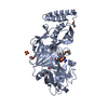
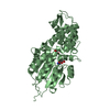
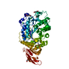
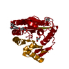
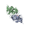
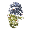
| Title | Crystal structure of human kappa class glutathione transferase in complex with S-hexylglutathione | ||||||
 Components Components | Glutathione S-transferase kappa 1 | ||||||
 Keywords Keywords | TRANSFERASE/TRANSFERASE INHIBITOR / kappa GST / Trx domain / GSH binding /  detoxification / GTX / glutathione transferase inhibitor complex / TRANSFERASE-TRANSFERASE INHIBITOR complex detoxification / GTX / glutathione transferase inhibitor complex / TRANSFERASE-TRANSFERASE INHIBITOR complex | ||||||
| Function / homology |  Function and homology informationGlutathione conjugation / glutathione peroxidase activity / peroxisomal matrix / glutathione transferase / glutathione transferase activity / glutathione metabolic process / epithelial cell differentiation / Peroxisomal protein import / peroxisome / mitochondrial matrix ...Glutathione conjugation / glutathione peroxidase activity / peroxisomal matrix / glutathione transferase / glutathione transferase activity / glutathione metabolic process / epithelial cell differentiation / Peroxisomal protein import / peroxisome / mitochondrial matrix / mitochondrion / extracellular exosome / membrane / cytosol Function and homology informationGlutathione conjugation / glutathione peroxidase activity / peroxisomal matrix / glutathione transferase / glutathione transferase activity / glutathione metabolic process / epithelial cell differentiation / Peroxisomal protein import / peroxisome / mitochondrial matrix ...Glutathione conjugation / glutathione peroxidase activity / peroxisomal matrix / glutathione transferase / glutathione transferase activity / glutathione metabolic process / epithelial cell differentiation / Peroxisomal protein import / peroxisome / mitochondrial matrix / mitochondrion / extracellular exosome / membrane / cytosolSimilarity search - Function | ||||||
| Biological species |  Homo sapiens (human) Homo sapiens (human) | ||||||
| Method | X-RAY DIFFRACTION / SYNCHROTRON / MOLECULAR REPLACEMENT / Resolution: 1.9 Å | ||||||
 Authors Authors | Wang, B. / Peng, Y. / Zhang, T. / Ding, J. | ||||||
 Citation Citation | Journal: Biochem.J. / Year: 2011 Title: Crystal structures and kinetic studies of human Kappa class glutathione transferase provide insights into the catalytic mechanism. Authors: Wang, B. / Peng, Y. / Zhang, T. / Ding, J. | ||||||
| History |
|
- Structure visualization
| Structure viewer | Molecule: MolmilJmol/JSmol |
|---|
- Downloads & links
-Download
| PDBx/mmCIF format | 3rpn.cif.gz | 557.3 KB | Display | PDBx/mmCIF format |
|---|---|---|---|---|
| PDB format | pdb3rpn.ent.gz | 461.6 KB | Display | PDB format |
| PDBx/mmJSON format | 3rpn.json.gz | Tree view | PDBx/mmJSON format | |
| Others |  Other downloads Other downloads |
-Validation report
| Arichive directory | https://data.pdbj.org/pub/pdb/validation_reports/rp/3rpnftp://data.pdbj.org/pub/pdb/validation_reports/rp/3rpn | HTTPS FTP |
|---|
-Related structure data
| Related structure data |  3rppC 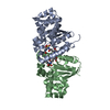 1yzxS C: citing same article ( S: Starting model for refinement |
|---|---|
| Similar structure data |


-Links
 PDBj
PDBj

- Assembly
| Deposited unit | 
| ||||||||
|---|---|---|---|---|---|---|---|---|---|
| 1 | 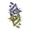
| ||||||||
| 2 | 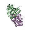
| ||||||||
| 3 | 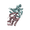
| ||||||||
| Unit cell |
|
-Components
| #1: Protein | Mass: 26600.016 Da / Num. of mol.: 6 Source method: isolated from a genetically manipulated source Source: (gene. exp.) Homo sapiens (human) / Gene: GSTK1, HDCMD47P / Plasmid: pET22b / Production host:  Escherichia coli (E. coli) / Strain (production host): BL21(DE3) / References: UniProt: Q9Y2Q3, glutathione transferase Escherichia coli (E. coli) / Strain (production host): BL21(DE3) / References: UniProt: Q9Y2Q3, glutathione transferase#2: Chemical | ChemComp-GTX /   Mass: 392.491 Da / Num. of mol.: 6 / Source method: obtained synthetically / Formula: C16H30N3O6S Mass: 392.491 Da / Num. of mol.: 6 / Source method: obtained synthetically / Formula: C16H30N3O6S#3: Water | ChemComp-HOH / | Water Mass: 18.015 Da / Num. of mol.: 804 / Source method: isolated from a natural source / Formula: H2O Mass: 18.015 Da / Num. of mol.: 804 / Source method: isolated from a natural source / Formula: H2O |
|---|
-Experimental details
-Experiment
| Experiment | Method: X-RAY DIFFRACTION / Number of used crystals: 1 |
|---|
- Sample preparation
| Crystal | Density Matthews: 2.49 Å3/Da / Density % sol: 50.6 % / Mosaicity: 0.527 ° |
|---|---|
| Crystal grow | Temperature: 293 K / Method: hanging drop / pH: 7 Details: 20% polyethylene glycol 3350, 0.2M NaSCN, pH 7.0, hanging drop, temperature 293K |
-Data collection
| Diffraction |
| |||||||||||||||||||||||||||||||||||||||||||||||||||||||||||||||||||||||||||||||||||||||||||||||||||
|---|---|---|---|---|---|---|---|---|---|---|---|---|---|---|---|---|---|---|---|---|---|---|---|---|---|---|---|---|---|---|---|---|---|---|---|---|---|---|---|---|---|---|---|---|---|---|---|---|---|---|---|---|---|---|---|---|---|---|---|---|---|---|---|---|---|---|---|---|---|---|---|---|---|---|---|---|---|---|---|---|---|---|---|---|---|---|---|---|---|---|---|---|---|---|---|---|---|---|---|---|
| Diffraction source | Source: SYNCHROTRON / Site: SSRF  / Beamline: BL17U / Beamline: BL17U | |||||||||||||||||||||||||||||||||||||||||||||||||||||||||||||||||||||||||||||||||||||||||||||||||||
| Detector | Type: ADSC QUANTUM 315 / Detector: CCD / Date: Jul 12, 2009 | |||||||||||||||||||||||||||||||||||||||||||||||||||||||||||||||||||||||||||||||||||||||||||||||||||
| Radiation | Protocol: SINGLE WAVELENGTH / Monochromatic (M) / Laue (L): M / Scattering type: x-ray | |||||||||||||||||||||||||||||||||||||||||||||||||||||||||||||||||||||||||||||||||||||||||||||||||||
| Radiation wavelength | Relative weight: 1 | |||||||||||||||||||||||||||||||||||||||||||||||||||||||||||||||||||||||||||||||||||||||||||||||||||
| Reflection | Resolution: 1.8→50 Å / Num. obs: 122112 / % possible obs: 73.3 % / Redundancy: 2.7 % / Rmerge(I) obs: 0.046 / Χ2: 1.128 / Net I/σ(I): 15.6 | |||||||||||||||||||||||||||||||||||||||||||||||||||||||||||||||||||||||||||||||||||||||||||||||||||
| Reflection shell |
|
- Processing
| Software |
| |||||||||||||||||||||||||||||||||||||||||||||||||||||||||||||||||||||||||||||
|---|---|---|---|---|---|---|---|---|---|---|---|---|---|---|---|---|---|---|---|---|---|---|---|---|---|---|---|---|---|---|---|---|---|---|---|---|---|---|---|---|---|---|---|---|---|---|---|---|---|---|---|---|---|---|---|---|---|---|---|---|---|---|---|---|---|---|---|---|---|---|---|---|---|---|---|---|---|---|
| Refinement | Method to determine structure: MOLECULAR REPLACEMENT Starting model: PDB ENTRY 1YZX Resolution: 1.9→29.951 Å / FOM work R set: 0.8341 / SU ML: 0.2 / σ(F): 0.55 / Phase error: 20.57 / Stereochemistry target values: ML
| |||||||||||||||||||||||||||||||||||||||||||||||||||||||||||||||||||||||||||||
| Solvent computation | Shrinkage radii: 1.17 Å / VDW probe radii: 1.4 Å / Solvent model: FLAT BULK SOLVENT MODEL / Bsol: 60.755 Å2 / ksol: 0.356 e/Å3 | |||||||||||||||||||||||||||||||||||||||||||||||||||||||||||||||||||||||||||||
| Displacement parameters |
| |||||||||||||||||||||||||||||||||||||||||||||||||||||||||||||||||||||||||||||
| Refinement step | Cycle: LAST / Resolution: 1.9→29.951 Å
| |||||||||||||||||||||||||||||||||||||||||||||||||||||||||||||||||||||||||||||
| Refine LS restraints |
| |||||||||||||||||||||||||||||||||||||||||||||||||||||||||||||||||||||||||||||
| LS refinement shell |
| |||||||||||||||||||||||||||||||||||||||||||||||||||||||||||||||||||||||||||||
| Refinement TLS params. | Method: refined / Origin x: 5.9704 Å / Origin y: -33.6106 Å / Origin z: 24.7373 Å
| |||||||||||||||||||||||||||||||||||||||||||||||||||||||||||||||||||||||||||||
| Refinement TLS group | Selection details: all |
