 Movie
Movie Controller

[English] 日本語
 Yorodumi
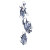
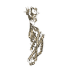
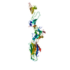
- PDB-3rkp: Crystal structure of BcpA*(D312A), the major pilin subunit of Bac... -
+ Open data

- Basic information
| Entry | Database: PDB / ID: 3rkp | ||||||
|---|---|---|---|---|---|---|---|
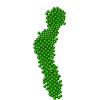
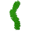
| Title | Crystal structure of BcpA*(D312A), the major pilin subunit of Bacillus cereus | ||||||
 Components Components | Collagen adhesion protein | ||||||
 Keywords Keywords |  CELL ADHESION / intramolecular amide bond / jelly-roll / Ig / pilin subunit CELL ADHESION / intramolecular amide bond / jelly-roll / Ig / pilin subunit | ||||||
| Function / homology |  Function and homology information Function and homology information | ||||||
| Biological species |  Bacillus cereus (bacteria) Bacillus cereus (bacteria) | ||||||
| Method | X-RAY DIFFRACTION / SYNCHROTRON / MOLECULAR REPLACEMENT / Resolution: 2.243 Å | ||||||
 Authors Authors | Hendrickx, A.P. / Poor, C.B. / Jureller, J.E. / Budzik, J.M. / He, C. / Schneewind, O. | ||||||
 Citation Citation | Journal: Mol.Microbiol. / Year: 2012 Title: Isopeptide bonds of the major pilin protein BcpA influence pilus structure and bundle formation on the surface of Bacillus cereus. Authors: Hendrickx, A.P. / Poor, C.B. / Jureller, J.E. / Budzik, J.M. / He, C. / Schneewind, O. | ||||||
| History |
|
- Structure visualization
| Structure viewer | Molecule: MolmilJmol/JSmol |
|---|
- Downloads & links
-Download
| PDBx/mmCIF format | 3rkp.cif.gz | 288 KB | Display | PDBx/mmCIF format |
|---|---|---|---|---|
| PDB format | pdb3rkp.ent.gz | 235.5 KB | Display | PDB format |
| PDBx/mmJSON format | 3rkp.json.gz | Tree view | PDBx/mmJSON format | |
| Others |  Other downloads Other downloads |
-Validation report
| Arichive directory | https://data.pdbj.org/pub/pdb/validation_reports/rk/3rkpftp://data.pdbj.org/pub/pdb/validation_reports/rk/3rkp | HTTPS FTP |
|---|
-Related structure data
| Related structure data |  3kptS S: Starting model for refinement |
|---|---|
| Similar structure data |


-Links
 PDBj
PDBj- Assembly
| Deposited unit | 
| ||||||||
|---|---|---|---|---|---|---|---|---|---|
| 1 | 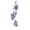
| ||||||||
| 2 | 
| ||||||||
| Unit cell |
|
-Components
| #1: Protein | Mass: 39233.578 Da / Num. of mol.: 2 / Fragment: unp residues 163-515 / Mutation: D312A, L182M, I261M, L357M, L426M Source method: isolated from a genetically manipulated source Source: (gene. exp.) Bacillus cereus (bacteria) / Strain: ATCC 14579 / DSM 31 / Gene: BC_2508 / Production host: Escherichia coli (E. coli) / References: UniProt: Q81D71#2: Water | ChemComp-HOH / | Water Mass: 18.015 Da / Num. of mol.: 133 / Source method: isolated from a natural source / Formula: H2O Mass: 18.015 Da / Num. of mol.: 133 / Source method: isolated from a natural source / Formula: H2O |
|---|
-Experimental details
-Experiment
| Experiment | Method: X-RAY DIFFRACTION / Number of used crystals: 1 |
|---|
- Sample preparation
| Crystal | Density Matthews: 2.77 Å3/Da / Density % sol: 55.56 % |
|---|---|
| Crystal grow | Temperature: 298 K / Method: vapor diffusion, hanging drop / pH: 7.7 Details: 0.1M HEPES pH 7.5 20% PEG 10,000, VAPOR DIFFUSION, HANGING DROP, temperature 298K |
-Data collection
| Diffraction | Mean temperature: 100 K |
|---|---|
| Diffraction source | Source: SYNCHROTRON / Site: APS  / Beamline: 23-ID-D / Wavelength: 1.0332 Å / Beamline: 23-ID-D / Wavelength: 1.0332 Å |
| Detector | Type: MAR CCD 130 mm / Detector: CCD / Date: Dec 20, 2010 |
| Radiation | Monochromator: Si(111) / Protocol: SINGLE WAVELENGTH / Monochromatic (M) / Laue (L): M / Scattering type: x-ray |
| Radiation wavelength | Wavelength: 1.0332 Å / Relative weight: 1 |
| Reflection | Resolution: 2.24→50 Å / Num. all: 41360 / Num. obs: 39085 / % possible obs: 94.5 % / Observed criterion σ(F): 2.24 / Observed criterion σ(I): 2.24 / Redundancy: 3.5 % / Rmerge(I) obs: 0.065 / Net I/σ(I): 16.96 |
| Reflection shell | Resolution: 2.24→2.28 Å / Rmerge(I) obs: 0.305 / Mean I/σ(I) obs: 2.68 / % possible all: 61.3 |
- Processing
| Software |
| |||||||||||||||||||||||||||||||||||||||||||||||||||||||||||||||||||||||||||||||||||||||||||||||||||||||||||||||||||||||||||||||||||||||||||||||||||||||||||||||||||||||||||||||
|---|---|---|---|---|---|---|---|---|---|---|---|---|---|---|---|---|---|---|---|---|---|---|---|---|---|---|---|---|---|---|---|---|---|---|---|---|---|---|---|---|---|---|---|---|---|---|---|---|---|---|---|---|---|---|---|---|---|---|---|---|---|---|---|---|---|---|---|---|---|---|---|---|---|---|---|---|---|---|---|---|---|---|---|---|---|---|---|---|---|---|---|---|---|---|---|---|---|---|---|---|---|---|---|---|---|---|---|---|---|---|---|---|---|---|---|---|---|---|---|---|---|---|---|---|---|---|---|---|---|---|---|---|---|---|---|---|---|---|---|---|---|---|---|---|---|---|---|---|---|---|---|---|---|---|---|---|---|---|---|---|---|---|---|---|---|---|---|---|---|---|---|---|---|---|---|---|
| Refinement | Method to determine structure: MOLECULAR REPLACEMENT Starting model: PDB ENTRY 3KPT Resolution: 2.243→38.781 Å / SU ML: 0.35 / σ(F): 0 / Phase error: 34.85 / Stereochemistry target values: ML
| |||||||||||||||||||||||||||||||||||||||||||||||||||||||||||||||||||||||||||||||||||||||||||||||||||||||||||||||||||||||||||||||||||||||||||||||||||||||||||||||||||||||||||||||
| Solvent computation | Shrinkage radii: 0.95 Å / VDW probe radii: 1.2 Å / Solvent model: FLAT BULK SOLVENT MODEL / Bsol: 39.213 Å2 / ksol: 0.324 e/Å3 | |||||||||||||||||||||||||||||||||||||||||||||||||||||||||||||||||||||||||||||||||||||||||||||||||||||||||||||||||||||||||||||||||||||||||||||||||||||||||||||||||||||||||||||||
| Displacement parameters |
| |||||||||||||||||||||||||||||||||||||||||||||||||||||||||||||||||||||||||||||||||||||||||||||||||||||||||||||||||||||||||||||||||||||||||||||||||||||||||||||||||||||||||||||||
| Refinement step | Cycle: LAST / Resolution: 2.243→38.781 Å
| |||||||||||||||||||||||||||||||||||||||||||||||||||||||||||||||||||||||||||||||||||||||||||||||||||||||||||||||||||||||||||||||||||||||||||||||||||||||||||||||||||||||||||||||
| Refine LS restraints |
| |||||||||||||||||||||||||||||||||||||||||||||||||||||||||||||||||||||||||||||||||||||||||||||||||||||||||||||||||||||||||||||||||||||||||||||||||||||||||||||||||||||||||||||||
| LS refinement shell |
| |||||||||||||||||||||||||||||||||||||||||||||||||||||||||||||||||||||||||||||||||||||||||||||||||||||||||||||||||||||||||||||||||||||||||||||||||||||||||||||||||||||||||||||||
| Refinement TLS params. | Method: refined / Refine-ID: X-RAY DIFFRACTION
| |||||||||||||||||||||||||||||||||||||||||||||||||||||||||||||||||||||||||||||||||||||||||||||||||||||||||||||||||||||||||||||||||||||||||||||||||||||||||||||||||||||||||||||||
| Refinement TLS group |
|
