 Movie
Movie Controller

[English] 日本語
 Yorodumi
- PDB-3rjz: X-ray crystal structure of the putative n-type atp pyrophosphatas... -
+ Open data

- Basic information
| Entry | Database: PDB / ID: 3rjz | |||||||||
|---|---|---|---|---|---|---|---|---|---|---|
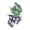
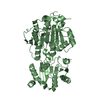
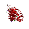
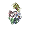
| Title | X-ray crystal structure of the putative n-type atp pyrophosphatase from pyrococcus furiosus, the northeast structural genomics target pfr23 | |||||||||
 Components Components | N-type ATP pyrophosphatase superfamily | |||||||||
 Keywords Keywords |  HYDROLASE / Structural Genomics / PSI-Biology / Northeast Structural Genomics Consortium / NESG / alpha-beta protein / n-type ATP pyrophosphatase HYDROLASE / Structural Genomics / PSI-Biology / Northeast Structural Genomics Consortium / NESG / alpha-beta protein / n-type ATP pyrophosphatase | |||||||||
| Function / homology |  Function and homology informationdiphthine-ammonia ligase activity / protein histidyl modification to diphthamide / ATP binding Function and homology informationdiphthine-ammonia ligase activity / protein histidyl modification to diphthamide / ATP bindingSimilarity search - Function | |||||||||
| Biological species |   Pyrococcus furiosus (archaea) Pyrococcus furiosus (archaea) | |||||||||
| Method | X-RAY DIFFRACTION / SYNCHROTRON / SAD / Resolution: 2.3 Å | |||||||||
 Authors Authors | Forouhar, F. / Lee, I. / Xiao, R. / Acton, T.B. / Montelione, G.T. / Hunt, J. / Tong, L. / Northeast Structural Genomics Consortium (NESG) | |||||||||
 Citation Citation | Journal: Acta Crystallogr.,Sect.F / Year: 2011 Title: A large conformational change in the putative ATP pyrophosphatase PF0828 induced by ATP binding. Authors: Forouhar, F. / Saadat, N. / Hussain, M. / Seetharaman, J. / Lee, I. / Janjua, H. / Xiao, R. / Shastry, R. / Acton, T.B. / Montelione, G.T. / Tong, L. | |||||||||
| History |
|
- Structure visualization
| Structure viewer | Molecule: MolmilJmol/JSmol |
|---|
- Downloads & links
-Download
| PDBx/mmCIF format | 3rjz.cif.gz | 51.5 KB | Display | PDBx/mmCIF format |
|---|---|---|---|---|
| PDB format | pdb3rjz.ent.gz | 40.4 KB | Display | PDB format |
| PDBx/mmJSON format | 3rjz.json.gz | Tree view | PDBx/mmJSON format | |
| Others |  Other downloads Other downloads |
-Validation report
| Arichive directory | https://data.pdbj.org/pub/pdb/validation_reports/rj/3rjzftp://data.pdbj.org/pub/pdb/validation_reports/rj/3rjz | HTTPS FTP |
|---|
-Related structure data


-Links
 PDBj
PDBj- Assembly
| Deposited unit | 
| ||||||||
|---|---|---|---|---|---|---|---|---|---|
| 1 | 
| ||||||||
| Unit cell |
|
-Components
| #1: Protein | Mass: 27107.328 Da / Num. of mol.: 1 Source method: isolated from a genetically manipulated source Source: (gene. exp.) Pyrococcus furiosus (archaea) / Strain: DSM 3638 / Gene: PF0828 / Plasmid: pET21 / Production host:  Escherichia coli (E. coli) / Strain (production host): BL21(DE3)+Magic / References: UniProt: Q8U2K6 Escherichia coli (E. coli) / Strain (production host): BL21(DE3)+Magic / References: UniProt: Q8U2K6 |
|---|---|
| #2: Water | ChemComp-HOH / Water Mass: 18.015 Da / Num. of mol.: 85 / Source method: isolated from a natural source / Formula: H2O Mass: 18.015 Da / Num. of mol.: 85 / Source method: isolated from a natural source / Formula: H2O |
-Experimental details
-Experiment
| Experiment | Method: X-RAY DIFFRACTION / Number of used crystals: 1 |
|---|
- Sample preparation
| Crystal | Density Matthews: 2.48 Å3/Da / Density % sol: 50.49 % |
|---|---|
| Crystal grow | Temperature: 277 K / Method: vapor diffusion, hanging drop / pH: 7.5 Details: Protein solution: 10 mM Tris (pH 7.5), 100 mM sodium chloride, 5 mM DTT, and 0.02% NaN3. Reservoir solution: 380 mM K/NaTartrate, , VAPOR DIFFUSION, HANGING DROP, temperature 277K |
-Data collection
| Diffraction | Mean temperature: 100 K |
|---|---|
| Diffraction source | Source: SYNCHROTRON / Site: NSLS  / Beamline: X4A / Wavelength: 0.92927 Å / Beamline: X4A / Wavelength: 0.92927 Å |
| Detector | Type: ADSC QUANTUM 4 / Detector: CCD / Date: Jun 16, 2003 / Details: mirrors |
| Radiation | Monochromator: Si 111 CHANNEL / Protocol: SINGLE WAVELENGTH / Monochromatic (M) / Laue (L): M / Scattering type: x-ray |
| Radiation wavelength | Wavelength: 0.92927 Å / Relative weight: 1 |
| Reflection | Resolution: 2.3→30 Å / Num. all: 23183 / Num. obs: 23091 / % possible obs: 99.6 % / Observed criterion σ(F): 0 / Observed criterion σ(I): 0 / Redundancy: 6.4 % / Biso Wilson estimate: 17.4 Å2 / Rmerge(I) obs: 0.082 / Rsym value: 0.072 / Net I/σ(I): 26.9 |
| Reflection shell | Resolution: 2.3→2.38 Å / Redundancy: 4.9 % / Rmerge(I) obs: 0.247 / Mean I/σ(I) obs: 6.1 / Num. unique all: 2300 / Rsym value: 0.22 / % possible all: 100 |
- Processing
| Software |
| |||||||||||||||||||||||||
|---|---|---|---|---|---|---|---|---|---|---|---|---|---|---|---|---|---|---|---|---|---|---|---|---|---|---|
| Refinement | Method to determine structure: SAD / Resolution: 2.3→19.93 Å / Rfactor Rfree error: 0.006 / Data cutoff high absF: 267007.46 / Data cutoff low absF: 0 / Isotropic thermal model: RESTRAINED / Cross valid method: THROUGHOUT / σ(F): 2 / σ(I): 2 / Stereochemistry target values: Engh & Huber
| |||||||||||||||||||||||||
| Solvent computation | Solvent model: FLAT MODEL / Bsol: 49.7501 Å2 / ksol: 0.4 e/Å3 | |||||||||||||||||||||||||
| Displacement parameters | Biso mean: 34.3 Å2
| |||||||||||||||||||||||||
| Refine analyze |
| |||||||||||||||||||||||||
| Refinement step | Cycle: LAST / Resolution: 2.3→19.93 Å
| |||||||||||||||||||||||||
| Refine LS restraints |
| |||||||||||||||||||||||||
| LS refinement shell | Resolution: 2.3→2.38 Å / Rfactor Rfree error: 0.024 / Total num. of bins used: 10
|
