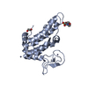
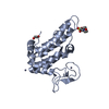
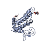
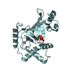
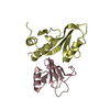
Hydrolase/DNA binding protein / alpha/beta domains / DNA repair / ATP Binding / DNA binding / nucleotide excision repair / Hydrolase-DNA binding protein complex
Function / homology
Function and homology information
transcription-coupled nucleotide-excision repair, DNA damage recognition / excinuclease ABC activity / excinuclease repair complex / RNA polymerase core enzyme binding / DNA translocase activity / nucleotide-excision repair, preincision complex assembly / DNA repair complex / SOS response / transcription-coupled nucleotide-excision repair / nucleotide-excision repair ...transcription-coupled nucleotide-excision repair, DNA damage recognition / excinuclease ABC activity / excinuclease repair complex / RNA polymerase core enzyme binding / DNA translocase activity / nucleotide-excision repair, preincision complex assembly / DNA repair complex / SOS response / transcription-coupled nucleotide-excision repair / nucleotide-excision repair / Hydrolases; Acting on acid anhydrides; Acting on acid anhydrides to facilitate cellular and subcellular movement / response to radiation / damaged DNA binding / hydrolase activity / DNA repair / DNA damage response / regulation of DNA-templated transcription / ATP hydrolysis activity / DNA binding / zinc ion binding / ATP binding / identical protein binding / cytosol Similarity search - Function
In the structure databanks used in Yorodumi, some data are registered as the other names, "COVID-19 virus" and "2019-nCoV". Here are the details of the virus and the list of structure data.
Jan 31, 2019. EMDB accession codes are about to change! (news from PDBe EMDB page)
EMDB accession codes are about to change! (news from PDBe EMDB page)
The allocation of 4 digits for EMDB accession codes will soon come to an end. Whilst these codes will remain in use, new EMDB accession codes will include an additional digit and will expand incrementally as the available range of codes is exhausted. The current 4-digit format prefixed with “EMD-” (i.e. EMD-XXXX) will advance to a 5-digit format (i.e. EMD-XXXXX), and so on. It is currently estimated that the 4-digit codes will be depleted around Spring 2019, at which point the 5-digit format will come into force.
The EM Navigator/Yorodumi systems omit the EMD- prefix.
Related info.:Q: What is EMD? / ID/Accession-code notation in Yorodumi/EM Navigator
Yorodumi is a browser for structure data from EMDB, PDB, SASBDB, etc.
This page is also the successor to EM Navigator detail page, and also detail information page/front-end page for Omokage search.
The word "yorodu" (or yorozu) is an old Japanese word meaning "ten thousand". "mi" (miru) is to see.
Related info.:EMDB / PDB / SASBDB / Comparison of 3 databanks / Yorodumi Search / Aug 31, 2016. New EM Navigator & Yorodumi / Yorodumi Papers / Jmol/JSmol / Function and homology information / Changes in new EM Navigator and Yorodumi
 Movie
Movie Controller


 Open data
 Basic information
 Components
 Keywords
 DNA repair /
  Function and homology information
 Authors
 Citation
 Structure visualization
 Downloads & links
 Other downloads


 PDBj
PDBj

 Assembly
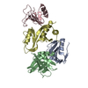

 Sample preparation
 / Beamline: 8.2.1 / Wavelength: 1 Å
 Processing