Method to determine structure: MOLECULAR REPLACEMENT / Resolution: 2.8→59.13 Å / Cor.coef. Fo:Fc: 0.934 / Cor.coef. Fo:Fc free: 0.881 / SU B: 17.593 / SU ML: 0.341 / Cross valid method: THROUGHOUT / ESU R Free: 0.414 / Stereochemistry target values: MAXIMUM LIKELIHOOD / Details: HYDROGENS HAVE BEEN USED IF PRESENT IN THE INPUT
Rfactor
Num. reflection
% reflection
Selection details
Rfree
0.28181
3845
5 %
RANDOM
Rwork
0.22274
-
-
-
obs
0.22574
73065
97.32 %
-
all
-
117208
-
-
Solvent computation
Ion probe radii: 0.8 Å / Shrinkage radii: 0.8 Å / VDW probe radii: 1.2 Å / Solvent model: BABINET MODEL WITH MASK
Displacement parameters
Biso mean: 60.62 Å2
Baniso -1
Baniso -2
Baniso -3
1-
0.18 Å2
0 Å2
0.16 Å2
2-
-
-0.24 Å2
0 Å2
3-
-
-
0.18 Å2
Refinement step
Cycle: LAST / Resolution: 2.8→59.13 Å
Protein
Nucleic acid
Ligand
Solvent
Total
Num. atoms
19068
0
10
41
19119
Refine LS restraints
Refine-ID
Type
Dev ideal
Dev ideal target
Number
X-RAY DIFFRACTION
r_bond_refined_d
0.007
0.022
19486
X-RAY DIFFRACTION
r_bond_other_d
X-RAY DIFFRACTION
r_angle_refined_deg
1.089
1.945
26600
X-RAY DIFFRACTION
r_angle_other_deg
X-RAY DIFFRACTION
r_dihedral_angle_1_deg
5.931
5
2502
X-RAY DIFFRACTION
r_dihedral_angle_2_deg
36.601
24.583
875
X-RAY DIFFRACTION
r_dihedral_angle_3_deg
16.603
15
2911
X-RAY DIFFRACTION
r_dihedral_angle_4_deg
16.505
15
104
X-RAY DIFFRACTION
r_chiral_restr
0.069
0.2
3006
X-RAY DIFFRACTION
r_gen_planes_refined
0.004
0.021
15082
X-RAY DIFFRACTION
r_gen_planes_other
X-RAY DIFFRACTION
r_nbd_refined
X-RAY DIFFRACTION
r_nbd_other
X-RAY DIFFRACTION
r_nbtor_refined
X-RAY DIFFRACTION
r_nbtor_other
X-RAY DIFFRACTION
r_xyhbond_nbd_refined
X-RAY DIFFRACTION
r_xyhbond_nbd_other
X-RAY DIFFRACTION
r_metal_ion_refined
X-RAY DIFFRACTION
r_metal_ion_other
X-RAY DIFFRACTION
r_symmetry_vdw_refined
X-RAY DIFFRACTION
r_symmetry_vdw_other
X-RAY DIFFRACTION
r_symmetry_hbond_refined
X-RAY DIFFRACTION
r_symmetry_hbond_other
X-RAY DIFFRACTION
r_symmetry_metal_ion_refined
X-RAY DIFFRACTION
r_symmetry_metal_ion_other
X-RAY DIFFRACTION
r_mcbond_it
X-RAY DIFFRACTION
r_mcbond_other
X-RAY DIFFRACTION
r_mcangle_it
X-RAY DIFFRACTION
r_scbond_it
X-RAY DIFFRACTION
r_scangle_it
X-RAY DIFFRACTION
r_rigid_bond_restr
X-RAY DIFFRACTION
r_sphericity_free
X-RAY DIFFRACTION
r_sphericity_bonded
Refine LS restraints NCS
Dom-ID: 1 / Refine-ID: X-RAY DIFFRACTION
Ens-ID
Auth asym-ID
Number
Type
Rms dev position (Å)
Weight position
1
A
1116
MEDIUMPOSITIONAL
0.23
0.5
1
A
905
LOOSEPOSITIONAL
0.33
5
1
A
1116
MEDIUMTHERMAL
5.29
2
1
A
905
LOOSETHERMAL
4.73
10
2
B
3100
MEDIUMPOSITIONAL
0.36
0.5
2
B
2810
LOOSEPOSITIONAL
0.49
5
2
B
3100
MEDIUMTHERMAL
5.1
2
2
B
2810
LOOSETHERMAL
5.39
10
3
C
803
MEDIUMPOSITIONAL
0.21
0.5
3
C
720
LOOSEPOSITIONAL
0.4
5
3
C
803
MEDIUMTHERMAL
3.99
2
3
C
720
LOOSETHERMAL
4.48
10
LS refinement shell
Resolution: 2.8→2.873 Å / Total num. of bins used: 20
Rfactor
Num. reflection
% reflection
Rfree
0.366
267
-
Rwork
0.341
5299
-
obs
-
-
98.44 %
+
About Yorodumi
-
News
-
Feb 9, 2022. New format data for meta-information of EMDB entries
New format data for meta-information of EMDB entries
Version 3 of the EMDB header file is now the official format.
The previous official version 1.9 will be removed from the archive.
In the structure databanks used in Yorodumi, some data are registered as the other names, "COVID-19 virus" and "2019-nCoV". Here are the details of the virus and the list of structure data.
Jan 31, 2019. EMDB accession codes are about to change! (news from PDBe EMDB page)
EMDB accession codes are about to change! (news from PDBe EMDB page)
The allocation of 4 digits for EMDB accession codes will soon come to an end. Whilst these codes will remain in use, new EMDB accession codes will include an additional digit and will expand incrementally as the available range of codes is exhausted. The current 4-digit format prefixed with “EMD-” (i.e. EMD-XXXX) will advance to a 5-digit format (i.e. EMD-XXXXX), and so on. It is currently estimated that the 4-digit codes will be depleted around Spring 2019, at which point the 5-digit format will come into force.
The EM Navigator/Yorodumi systems omit the EMD- prefix.
Related info.:Q: What is EMD? / ID/Accession-code notation in Yorodumi/EM Navigator
Yorodumi is a browser for structure data from EMDB, PDB, SASBDB, etc.
This page is also the successor to EM Navigator detail page, and also detail information page/front-end page for Omokage search.
The word "yorodu" (or yorozu) is an old Japanese word meaning "ten thousand". "mi" (miru) is to see.
Related info.:EMDB / PDB / SASBDB / Comparison of 3 databanks / Yorodumi Search / Aug 31, 2016. New EM Navigator & Yorodumi / Yorodumi Papers / Jmol/JSmol / Function and homology information / Changes in new EM Navigator and Yorodumi
 Movie
Movie Controller

 Yorodumi
 Open data
 Basic information
 Components
 Keywords
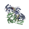
 beta-barrel /
  Function and homology information
 Authors
 Citation
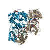
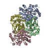
 Structure visualization
 Downloads & links
 Other downloads


 PDBj
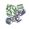
PDBj Assembly
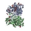
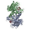
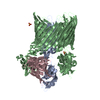


 Mass: 96.063 Da / Num. of mol.: 2 / Source method: obtained synthetically / Formula: SO4
 Mass: 18.015 Da / Num. of mol.: 41 / Source method: isolated from a natural source / Formula: H2O
 Sample preparation
 / Beamline: ID23-1 / Wavelength: 1.0723 Å
 Processing