 Movie
Movie Controller

[English] 日本語
 Yorodumi
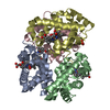
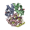
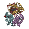
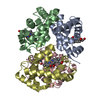
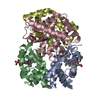
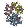
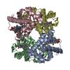
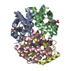
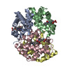
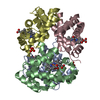
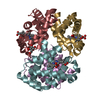
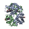
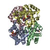
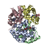
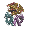
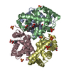
- PDB-3r5i: Crystal structure of liganded Hemoglobin complexed with a potent ... -
+ Open data

- Basic information
| Entry | Database: PDB / ID: 3r5i | ||||||
|---|---|---|---|---|---|---|---|
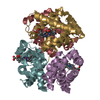
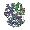
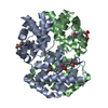
| Title | Crystal structure of liganded Hemoglobin complexed with a potent Antisickling agent, INN-312 | ||||||
 Components Components | (Hemoglobin subunit ... ) x 2 ) x 2 | ||||||
 Keywords Keywords | oxygen transport / oxygen binding / Heme protein / Antisickling / globin fold / red blood cell | ||||||
| Function / homology |  Function and homology information Function and homology informationcellular oxidant detoxification / nitric oxide transport / hemoglobin alpha binding / haptoglobin-hemoglobin complex / organic acid binding / hemoglobin binding / renal absorption / hemoglobin complex / oxygen transport / Scavenging of heme from plasma ...cellular oxidant detoxification / nitric oxide transport / hemoglobin alpha binding / haptoglobin-hemoglobin complex / organic acid binding / hemoglobin binding / renal absorption / hemoglobin complex / oxygen transport / Scavenging of heme from plasma / endocytic vesicle lumen / blood vessel diameter maintenance / hydrogen peroxide catabolic process / oxygen carrier activity / Late endosomal microautophagy / Heme signaling / carbon dioxide transport / Erythrocytes take up oxygen and release carbon dioxide / response to hydrogen peroxide / Erythrocytes take up carbon dioxide and release oxygen / Cytoprotection by HMOX1 / platelet aggregation / oxygen binding / regulation of blood pressure / Chaperone Mediated Autophagy / positive regulation of nitric oxide biosynthetic process / tertiary granule lumen / Factors involved in megakaryocyte development and platelet production / ficolin-1-rich granule lumen / blood microparticle / iron ion binding / heme binding / Neutrophil degranulation / extracellular space / extracellular exosome / extracellular region / membrane / metal ion binding / cytosolSimilarity search - Function | ||||||
| Biological species |  Homo sapiens (human) Homo sapiens (human) | ||||||
| Method | X-RAY DIFFRACTION / isomorphous refinement / Resolution: 2.2 Å | ||||||
 Authors Authors | Safo, M.K. / Musayev, F.N. / Safo, R.P. / Daniels, D. / Eseonu, D.N. / Parra, J. | ||||||
 Citation Citation | Journal: To be Published Title: Structural and in Vitro Chracterization of Pyridyl Derivatives of Benzaldehydes : Highly Potent Antisickling Agents Authors: Abulmalik, O. / Ghatge, M.S. / Nnamani, I. / Eseonu, D.N. / Musayev, F.N. / Danso-Danquah, R. / Venitz, J. / Asakura, T. / Abraham, D.J. / Safo, M.K. | ||||||
| History |
|
- Structure visualization
| Structure viewer | Molecule: MolmilJmol/JSmol |
|---|
- Downloads & links
-Download
| PDBx/mmCIF format | 3r5i.cif.gz | 132.7 KB | Display | PDBx/mmCIF format |
|---|---|---|---|---|
| PDB format | pdb3r5i.ent.gz | 104.8 KB | Display | PDB format |
| PDBx/mmJSON format | 3r5i.json.gz | Tree view | PDBx/mmJSON format | |
| Others |  Other downloads Other downloads |
-Validation report
| Arichive directory | https://data.pdbj.org/pub/pdb/validation_reports/r5/3r5iftp://data.pdbj.org/pub/pdb/validation_reports/r5/3r5i | HTTPS FTP |
|---|
-Related structure data
| Related structure data |  3ic0S S: Starting model for refinement |
|---|---|
| Similar structure data |


-Links
 PDBj
PDBj


- Assembly
| Deposited unit | 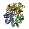
| ||||||||
|---|---|---|---|---|---|---|---|---|---|
| 1 |
| ||||||||
| Unit cell |
|
-Components
-Hemoglobin subunit ... , 2 types, 4 molecules ACBD
| #1: Protein | / Alpha-globin / Hemoglobin alpha chain Mass: 15150.353 Da / Num. of mol.: 2 / Source method: isolated from a natural source / Source: (natural) Homo sapiens (human) / Cell: red blood cells / References: UniProt: P69905#2: Protein | / Beta-globin / Hemoglobin beta chain / LVV-hemorphin-7Mass: 15890.198 Da / Num. of mol.: 2 / Source method: isolated from a natural source / Source: (natural) Homo sapiens (human) / Cell: red blood cells / References: UniProt: P68871 |
|---|
-Non-polymers , 5 types, 323 molecules 


| #3: Chemical | ChemComp-OXY / Oxygen Mass: 31.999 Da / Num. of mol.: 4 / Source method: obtained synthetically / Formula: O2 Mass: 31.999 Da / Num. of mol.: 4 / Source method: obtained synthetically / Formula: O2#4: Chemical | ChemComp-HEM / Heme B Mass: 616.487 Da / Num. of mol.: 4 / Source method: obtained synthetically / Formula: C34H32FeN4O4 Mass: 616.487 Da / Num. of mol.: 4 / Source method: obtained synthetically / Formula: C34H32FeN4O4#5: Chemical |  Mass: 243.258 Da / Num. of mol.: 3 / Source method: obtained synthetically / Formula: C14H13NO3 Mass: 243.258 Da / Num. of mol.: 3 / Source method: obtained synthetically / Formula: C14H13NO3#6: Chemical | Sulfate Mass: 96.063 Da / Num. of mol.: 3 / Source method: obtained synthetically / Formula: SO4 Mass: 96.063 Da / Num. of mol.: 3 / Source method: obtained synthetically / Formula: SO4#7: Water | ChemComp-HOH / | WaterMass: 18.015 Da / Num. of mol.: 309 / Source method: isolated from a natural source / Formula: H2O |
|---|
-Experimental details
-Experiment
| Experiment | Method: X-RAY DIFFRACTION / Number of used crystals: 1 |
|---|
- Sample preparation
| Crystal | Density Matthews: 2.84 Å3/Da / Density % sol: 56.62 % |
|---|---|
| Crystal grow | Temperature: 298 K / Method: liquid diffusion / pH: 6.4 Details: 3.2 M Na+/K+ phosphate, pH 6.4, LIQUID DIFFUSION, temperature 298K |
-Data collection
| Diffraction | Mean temperature: 100 K |
|---|---|
| Diffraction source | Source: ROTATING ANODE / Type: RIGAKU MICROMAX-007 / Wavelength: 1.54 Å |
| Detector | Type: RIGAKU RAXIS / Detector: IMAGE PLATE / Date: Jan 23, 2011 / Details: mirrors |
| Radiation | Protocol: SINGLE WAVELENGTH / Scattering type: x-ray |
| Radiation wavelength | Wavelength: 1.54 Å / Relative weight: 1 |
| Reflection | Resolution: 2.2→29.51 Å / Num. all: 36347 / Num. obs: 36347 / % possible obs: 99.6 % / Observed criterion σ(F): 0 / Observed criterion σ(I): 0 / Redundancy: 4.61 % / Biso Wilson estimate: 50 Å2 / Rmerge(I) obs: 0.067 / Rsym value: 0.075 / Net I/σ(I): 10.5 |
| Reflection shell | Resolution: 2.2→2.28 Å / Redundancy: 4.59 % / Rmerge(I) obs: 0.342 / Mean I/σ(I) obs: 3.3 / Num. unique all: 3612 / Rsym value: 0.387 / % possible all: 100 |
- Processing
| Software |
| ||||||||||||||||||||||||||||
|---|---|---|---|---|---|---|---|---|---|---|---|---|---|---|---|---|---|---|---|---|---|---|---|---|---|---|---|---|---|
| Refinement | Method to determine structure: isomorphous refinement Starting model: PDB entry 3IC0 Resolution: 2.2→20.09 Å / Rfactor Rfree error: 0.006 / Occupancy max: 1 / Occupancy min: 1 / Data cutoff high absF: 2084350 / Data cutoff low absF: 0 / Isotropic thermal model: RESTRAINED / Cross valid method: THROUGHOUT / σ(F): 0 / Stereochemistry target values: Engh & Huber
| ||||||||||||||||||||||||||||
| Solvent computation | Solvent model: FLAT MODEL / Bsol: 72.0694 Å2 / ksol: 0.3784 e/Å3 | ||||||||||||||||||||||||||||
| Displacement parameters | Biso max: 100 Å2 / Biso mean: 46.4788 Å2 / Biso min: 23.92 Å2
| ||||||||||||||||||||||||||||
| Refine analyze |
| ||||||||||||||||||||||||||||
| Refinement step | Cycle: LAST / Resolution: 2.2→20.09 Å
| ||||||||||||||||||||||||||||
| Refine LS restraints |
| ||||||||||||||||||||||||||||
| LS refinement shell | Resolution: 2.2→2.3 Å / Rfactor Rfree error: 0.025 / Total num. of bins used: 8
| ||||||||||||||||||||||||||||
| Xplor file |
|
