Resolution: 2.2→30 Å / Cor.coef. Fo:Fc: 0.953 / Cor.coef. Fo:Fc free: 0.911 / SU B: 10.249 / SU ML: 0.142 / TLS residual ADP flag: LIKELY RESIDUAL / Isotropic thermal model: Isotropic / Cross valid method: THROUGHOUT / σ(F): 0 / ESU R: 0.283 / ESU R Free: 0.218 / Stereochemistry target values: Engh & Huber Details: Simulated annealing was performed with the CNS software in the first cycle or refinement. Used TLS refinement. Tyr 145 from the B and D molecules presented weak density for an alternate ...Details: Simulated annealing was performed with the CNS software in the first cycle or refinement. Used TLS refinement. Tyr 145 from the B and D molecules presented weak density for an alternate conformation that was not modeled.
Rfactor
Num. reflection
% reflection
Selection details
Rfree
0.23266
1530
5.1 %
RANDOM
Rwork
0.165
-
-
-
all
0.1685
30163
-
-
obs
0.1685
28633
99.91 %
-
Solvent computation
Ion probe radii: 0.8 Å / Shrinkage radii: 0.8 Å / VDW probe radii: 1.2 Å / Solvent model: BABINET MODEL WITH MASK
Displacement parameters
Biso mean: 27.571 Å2
Baniso -1
Baniso -2
Baniso -3
1-
-1 Å2
0 Å2
0 Å2
2-
-
-0.15 Å2
0 Å2
3-
-
-
1.15 Å2
Refinement step
Cycle: LAST / Resolution: 2.2→30 Å
Protein
Nucleic acid
Ligand
Solvent
Total
Num. atoms
4395
0
182
443
5020
Refine LS restraints
Refine-ID
Type
Dev ideal
Dev ideal target
Number
X-RAY DIFFRACTION
r_bond_refined_d
0.015
0.022
4716
X-RAY DIFFRACTION
r_angle_refined_deg
1.399
2.065
6468
X-RAY DIFFRACTION
r_dihedral_angle_1_deg
5.525
5
571
X-RAY DIFFRACTION
r_dihedral_angle_2_deg
36.13
24.341
182
X-RAY DIFFRACTION
r_dihedral_angle_3_deg
14.431
15
729
X-RAY DIFFRACTION
r_dihedral_angle_4_deg
12.223
15
10
X-RAY DIFFRACTION
r_chiral_restr
0.092
0.2
705
X-RAY DIFFRACTION
r_gen_planes_refined
0.006
0.02
3548
X-RAY DIFFRACTION
r_nbd_refined
0.216
0.2
3065
X-RAY DIFFRACTION
r_nbtor_refined
0.312
0.2
3314
X-RAY DIFFRACTION
r_xyhbond_nbd_refined
0.161
0.2
588
X-RAY DIFFRACTION
r_metal_ion_refined
0.025
0.2
2
X-RAY DIFFRACTION
r_symmetry_vdw_refined
0.151
0.2
58
X-RAY DIFFRACTION
r_symmetry_hbond_refined
0.156
0.2
15
X-RAY DIFFRACTION
r_mcbond_it
0.707
1.5
2886
X-RAY DIFFRACTION
r_mcangle_it
1.196
2
4576
X-RAY DIFFRACTION
r_scbond_it
2.048
3
2061
X-RAY DIFFRACTION
r_scangle_it
2.996
4.5
1884
LS refinement shell
Resolution: 2.2→2.257 Å / Total num. of bins used: 20
Rfactor
Num. reflection
% reflection
Rfree
0.274
108
-
Rwork
0.168
2039
-
obs
-
2039
99.26 %
Refinement TLS params.
Method: refined / Refine-ID: X-RAY DIFFRACTION
ID
L11 (°2)
L12 (°2)
L13 (°2)
L22 (°2)
L23 (°2)
L33 (°2)
S11 (Å °)
S12 (Å °)
S13 (Å °)
S21 (Å °)
S22 (Å °)
S23 (Å °)
S31 (Å °)
S32 (Å °)
S33 (Å °)
T11 (Å2)
T12 (Å2)
T13 (Å2)
T22 (Å2)
T23 (Å2)
T33 (Å2)
Origin x (Å)
Origin y (Å)
Origin z (Å)
1
3.0878
0.0346
0.0893
1.2604
0.2159
1.4903
-0.0242
0.0197
-0.0627
0.0231
0.0349
0.042
0.1783
0.1128
-0.0107
-0.0976
0.0092
0.0074
-0.1415
0.0019
-0.1875
-14.725
-6.8243
-4.3671
2
4.068
-0.2974
-1.2965
1.8756
-0.1522
2.2526
0.0621
-0.0077
0.6078
0.1159
0.063
-0.05
-0.3672
-0.2119
-0.1251
-0.0672
0.0291
-0.0393
-0.1102
0.0067
-0.0144
-31.6549
9.3321
-0.0934
3
3.397
0.3197
0.497
1.7903
0.4025
1.588
-0.0199
-0.032
-0.2864
-0.1146
-0.0218
-0.1375
0.1674
-0.0991
0.0417
-0.1076
-0.0177
0.0376
-0.1571
0.0152
-0.1444
-27.9532
-11.6629
23.8805
4
2.7702
0.1424
-1.1426
1.9285
0.8494
2.5942
0.0295
0.0123
0.2906
-0.1109
0.0139
-0.0652
-0.2953
0.1211
-0.0433
-0.116
-0.0116
-0.0279
-0.1212
-0.0061
-0.1331
-11.2152
5.1936
25.1298
Refinement TLS group
ID
Refine-ID
Refine TLS-ID
Auth asym-ID
Label asym-ID
Auth seq-ID
Label seq-ID
1
X-RAY DIFFRACTION
1
A
A
1 - 141
1 - 141
2
X-RAY DIFFRACTION
1
A
G
200
1
3
X-RAY DIFFRACTION
2
B
B
2 - 145
2 - 145
4
X-RAY DIFFRACTION
2
B
H
200
1
5
X-RAY DIFFRACTION
3
C
C
1 - 141
1 - 141
6
X-RAY DIFFRACTION
3
C
I
200
1
7
X-RAY DIFFRACTION
4
D
D
1 - 145
1 - 145
8
X-RAY DIFFRACTION
4
D
J
200
1
+
About Yorodumi
-
News
-
Feb 9, 2022. New format data for meta-information of EMDB entries
New format data for meta-information of EMDB entries
Version 3 of the EMDB header file is now the official format.
The previous official version 1.9 will be removed from the archive.
In the structure databanks used in Yorodumi, some data are registered as the other names, "COVID-19 virus" and "2019-nCoV". Here are the details of the virus and the list of structure data.
Jan 31, 2019. EMDB accession codes are about to change! (news from PDBe EMDB page)
EMDB accession codes are about to change! (news from PDBe EMDB page)
The allocation of 4 digits for EMDB accession codes will soon come to an end. Whilst these codes will remain in use, new EMDB accession codes will include an additional digit and will expand incrementally as the available range of codes is exhausted. The current 4-digit format prefixed with “EMD-” (i.e. EMD-XXXX) will advance to a 5-digit format (i.e. EMD-XXXXX), and so on. It is currently estimated that the 4-digit codes will be depleted around Spring 2019, at which point the 5-digit format will come into force.
The EM Navigator/Yorodumi systems omit the EMD- prefix.
Related info.:Q: What is EMD? / ID/Accession-code notation in Yorodumi/EM Navigator
Yorodumi is a browser for structure data from EMDB, PDB, SASBDB, etc.
This page is also the successor to EM Navigator detail page, and also detail information page/front-end page for Omokage search.
The word "yorodu" (or yorozu) is an old Japanese word meaning "ten thousand". "mi" (miru) is to see.
Related info.:EMDB / PDB / SASBDB / Comparison of 3 databanks / Yorodumi Search / Aug 31, 2016. New EM Navigator & Yorodumi / Yorodumi Papers / Jmol/JSmol / Function and homology information / Changes in new EM Navigator and Yorodumi
 Movie
Movie Controller

 Yorodumi
 Open data
 Basic information
 Components
 Keywords
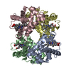
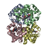
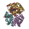
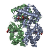
 hemoglobin / aquomet / Cerdocyon thous / OXYGEN STORAGE-TRANSPORT COMPLEX
 Function and homology information
 Authors
 Citation
 Structure visualization
 Downloads & links
 Other downloads


 PDBj
PDBj


 Assembly

 Mass: 96.063 Da / Num. of mol.: 2 / Source method: obtained synthetically / Formula: SO4
 Mass: 616.487 Da / Num. of mol.: 4 / Source method: obtained synthetically / Formula: C34H32FeN4O4
 Mass: 18.015 Da / Num. of mol.: 443 / Source method: isolated from a natural source / Formula: H2O
 Sample preparation
 / Beamline: D03B-MX1 / Wavelength: 1.427 Å
 Processing