 Movie
Movie Controller

[English] 日本語
 Yorodumi
- PDB-3r3j: Kinetic and structural characterization of Plasmodium falciparum ... -
+ Open data

- Basic information
| Entry | Database: PDB / ID: 3r3j | ||||||
|---|---|---|---|---|---|---|---|
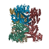
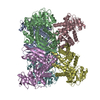
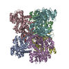
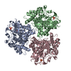
| Title | Kinetic and structural characterization of Plasmodium falciparum glutamate dehydrogenase 2 | ||||||
 Components Components | Glutamate dehydrogenase | ||||||
 Keywords Keywords | OXIDOREDUCTASE / Rossmann Fold / Apicoplast / Plasmodium falciparum | ||||||
| Function / homology |  Function and homology informationapicoplast / glutamate biosynthetic process / glutamate dehydrogenase (NADP+) activity / amino acid metabolic process / nucleotide binding / cytosol Function and homology informationapicoplast / glutamate biosynthetic process / glutamate dehydrogenase (NADP+) activity / amino acid metabolic process / nucleotide binding / cytosolSimilarity search - Function | ||||||
| Biological species |  Plasmodium falciparum (malaria parasite P. falciparum) Plasmodium falciparum (malaria parasite P. falciparum) | ||||||
| Method | X-RAY DIFFRACTION / SYNCHROTRON / MOLECULAR REPLACEMENT / Resolution: 3.1 Å | ||||||
 Authors Authors | Zocher, K. / Fritz-Wolf, K. / Kehr, S. / Rahlfs, S. / Becker, K. | ||||||
 Citation Citation | Journal: Mol.Biochem.Parasitol. / Year: 2012 Title: Biochemical and structural characterization of Plasmodium falciparum glutamate dehydrogenase 2. Authors: Zocher, K. / Fritz-Wolf, K. / Kehr, S. / Fischer, M. / Rahlfs, S. / Becker, K. | ||||||
| History |
|
- Structure visualization
| Structure viewer | Molecule: MolmilJmol/JSmol |
|---|
- Downloads & links
-Download
| PDBx/mmCIF format | 3r3j.cif.gz | 520.7 KB | Display | PDBx/mmCIF format |
|---|---|---|---|---|
| PDB format | pdb3r3j.ent.gz | 431.8 KB | Display | PDB format |
| PDBx/mmJSON format | 3r3j.json.gz | Tree view | PDBx/mmJSON format | |
| Others |  Other downloads Other downloads |
-Validation report
| Arichive directory | https://data.pdbj.org/pub/pdb/validation_reports/r3/3r3jftp://data.pdbj.org/pub/pdb/validation_reports/r3/3r3j | HTTPS FTP |
|---|
-Related structure data
| Related structure data |  2bmaS S: Starting model for refinement |
|---|---|
| Similar structure data |


-Links
 PDBj
PDBj- Assembly
| Deposited unit | 
| ||||||||
|---|---|---|---|---|---|---|---|---|---|
| 1 |
| ||||||||
| Unit cell |
|
-Components
| #1: Protein | Mass: 51261.422 Da / Num. of mol.: 6 / Fragment: UNP Residues 55-510 / Mutation: T161A Source method: isolated from a genetically manipulated source Source: (gene. exp.) Plasmodium falciparum (malaria parasite P. falciparum)Strain: 3D7 / Gene: PF14_0286 / Plasmid: pET28a / Production host:  Escherichia coli (E. coli) / Strain (production host): KRX Escherichia coli (E. coli) / Strain (production host): KRXReferences: UniProt: Q8ILF7, glutamate dehydrogenase [NAD(P)+] #2: Water | ChemComp-HOH / | Water Mass: 18.015 Da / Num. of mol.: 19 / Source method: isolated from a natural source / Formula: H2O Mass: 18.015 Da / Num. of mol.: 19 / Source method: isolated from a natural source / Formula: H2O |
|---|
-Experimental details
-Experiment
| Experiment | Method: X-RAY DIFFRACTION / Number of used crystals: 1 |
|---|
- Sample preparation
| Crystal | Density Matthews: 2.86 Å3/Da / Density % sol: 56.97 % |
|---|---|
| Crystal grow | Temperature: 293 K / Method: vapor diffusion, hanging drop / pH: 8.5 Details: 0.1M TRIS, 0.2M magnesium chloride hexahydrate, 30% PEG 4000, 0.01M spermine tetra HCl, pH 8.5, VAPOR DIFFUSION, HANGING DROP, temperature 293K |
-Data collection
| Diffraction | Mean temperature: 100 K |
|---|---|
| Diffraction source | Source: SYNCHROTRON / Site: SLS  / Beamline: X10SA / Wavelength: 0.97932 Å / Beamline: X10SA / Wavelength: 0.97932 Å |
| Detector | Type: MARMOSAIC 225 mm CCD / Detector: CCD / Date: May 30, 2009 |
| Radiation | Protocol: SINGLE WAVELENGTH / Monochromatic (M) / Laue (L): M / Scattering type: x-ray |
| Radiation wavelength | Wavelength: 0.97932 Å / Relative weight: 1 |
| Reflection | Resolution: 3.1→25 Å / Num. all: 64633 / Num. obs: 64381 / % possible obs: 99.6 % / Observed criterion σ(F): 0 / Observed criterion σ(I): -3 / Redundancy: 10.5 % / Biso Wilson estimate: 42.7 Å2 / Rsym value: 0.15 / Net I/σ(I): 15.3 |
| Reflection shell | Resolution: 3.1→3.2 Å / Redundancy: 9.2 % / Rmerge(I) obs: 0.78 / Mean I/σ(I) obs: 2.5 / Num. unique all: 5744 / % possible all: 99.5 |
- Processing
| Software |
| ||||||||||||||||||||||||||||||||||||
|---|---|---|---|---|---|---|---|---|---|---|---|---|---|---|---|---|---|---|---|---|---|---|---|---|---|---|---|---|---|---|---|---|---|---|---|---|---|
| Refinement | Method to determine structure: MOLECULAR REPLACEMENT Starting model: PDB ENTRY 2BMA Resolution: 3.1→24.99 Å / Rfactor Rfree error: 0.005 / Data cutoff high absF: 5503951.09 / Data cutoff low absF: 0 / Isotropic thermal model: RESTRAINED / Cross valid method: THROUGHOUT / σ(F): 0 / Details: BULK SOLVENT MODEL USED
| ||||||||||||||||||||||||||||||||||||
| Solvent computation | Solvent model: FLAT MODEL / Bsol: 44.4999 Å2 / ksol: 0.35 e/Å3 | ||||||||||||||||||||||||||||||||||||
| Displacement parameters | Biso mean: 72 Å2
| ||||||||||||||||||||||||||||||||||||
| Refine analyze |
| ||||||||||||||||||||||||||||||||||||
| Refinement step | Cycle: LAST / Resolution: 3.1→24.99 Å
| ||||||||||||||||||||||||||||||||||||
| Refine LS restraints |
| ||||||||||||||||||||||||||||||||||||
| Refine LS restraints NCS | NCS model details: CONSTR | ||||||||||||||||||||||||||||||||||||
| LS refinement shell | Resolution: 3.1→3.29 Å / Rfactor Rfree error: 0.015 / Total num. of bins used: 6
| ||||||||||||||||||||||||||||||||||||
| Xplor file |
|
