 Movie
Movie Controller

[English] 日本語
 Yorodumi
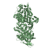
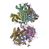
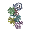
- PDB-3r2x: Crystal structure of the de novo designed binding protein HB36.3 ... -
+ Open data

- Basic information
| Entry | Database: PDB / ID: 3r2x | |||||||||
|---|---|---|---|---|---|---|---|---|---|---|
| Title | Crystal structure of the de novo designed binding protein HB36.3 in complex the the 1918 influenza virus hemagglutinin | |||||||||
 Components Components |
| |||||||||
 Keywords Keywords |  VIRAL PROTEIN/DE NOVO PROTEIN / hemagglutinin / glycoprotein / VIRAL PROTEIN-DE NOVO PROTEIN complex VIRAL PROTEIN/DE NOVO PROTEIN / hemagglutinin / glycoprotein / VIRAL PROTEIN-DE NOVO PROTEIN complex | |||||||||
| Function / homology |  Function and homology informationviral budding from plasma membrane / clathrin-dependent endocytosis of virus by host cell / host cell surface receptor binding / fusion of virus membrane with host plasma membrane / fusion of virus membrane with host endosome membrane / viral envelope / virion attachment to host cell / host cell plasma membrane / virion membrane / membrane Function and homology informationviral budding from plasma membrane / clathrin-dependent endocytosis of virus by host cell / host cell surface receptor binding / fusion of virus membrane with host plasma membrane / fusion of virus membrane with host endosome membrane / viral envelope / virion attachment to host cell / host cell plasma membrane / virion membrane / membraneSimilarity search - Function | |||||||||
| Biological species |   Influenza A virus Influenza A virusartificial gene (others) | |||||||||
| Method | X-RAY DIFFRACTION / SYNCHROTRON / MOLECULAR REPLACEMENT / Resolution: 3.1 Å | |||||||||
 Authors Authors | Ekiert, D.C. / Wilson, I.A. | |||||||||
 Citation Citation | Journal: Science / Year: 2011 Title: Computational design of proteins targeting the conserved stem region of influenza hemagglutinin. Authors: Fleishman, S.J. / Whitehead, T.A. / Ekiert, D.C. / Dreyfus, C. / Corn, J.E. / Strauch, E.M. / Wilson, I.A. / Baker, D. | |||||||||
| History |
|
- Structure visualization
| Structure viewer | Molecule: MolmilJmol/JSmol |
|---|
- Downloads & links
-Download
| PDBx/mmCIF format | 3r2x.cif.gz | 244.2 KB | Display | PDBx/mmCIF format |
|---|---|---|---|---|
| PDB format | pdb3r2x.ent.gz | 204.8 KB | Display | PDB format |
| PDBx/mmJSON format | 3r2x.json.gz | Tree view | PDBx/mmJSON format | |
| Others |  Other downloads Other downloads |
-Validation report
| Arichive directory | https://data.pdbj.org/pub/pdb/validation_reports/r2/3r2xftp://data.pdbj.org/pub/pdb/validation_reports/r2/3r2x | HTTPS FTP |
|---|
-Related structure data
| Similar structure data |
|---|


-Links
 PDBj
PDBj


- Assembly
| Deposited unit | 
| ||||||||
|---|---|---|---|---|---|---|---|---|---|
| 1 | 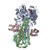
| ||||||||
| Unit cell |
|
-Components
| #1: Protein | / 1918 hemagglutinin receptor binding domain Mass: 36452.797 Da / Num. of mol.: 1 / Fragment: HA1 chain (UNP residues 18-344) Source method: isolated from a genetically manipulated source Source: (gene. exp.) Influenza A virus / Strain: A/South Carolina/1/1918 (H1N1) / Gene: HA, hemagglutinin / Production host:  Trichoplusia ni (cabbage looper) / Strain (production host): High Five / References: UniProt: Q9WFX3 Trichoplusia ni (cabbage looper) / Strain (production host): High Five / References: UniProt: Q9WFX3 |
|---|---|
| #2: Protein | / 1918 hemagglutinin fusion subunit Mass: 20394.445 Da / Num. of mol.: 1 / Fragment: HA2 chain (UNP residues 345-520) Source method: isolated from a genetically manipulated source Source: (gene. exp.) Influenza A virus / Strain: A/South Carolina/1/1918 (H1N1) / Gene: HA, hemagglutinin / Production host: Trichoplusia ni (cabbage looper) / Strain (production host): High Five / References: UniProt: Q9WFX3 |
| #3: Protein | Mass: 10605.185 Da / Num. of mol.: 1 Source method: isolated from a genetically manipulated source Source: (gene. exp.) artificial gene (others) / Production host:  Escherichia coli (E. coli) / Strain (production host): Rosetta 2 (BL21/DE3) Escherichia coli (E. coli) / Strain (production host): Rosetta 2 (BL21/DE3) |
| #4: Polysaccharide | alpha-D-mannopyranose-(1-3)-beta-D-mannopyranose-(1-4)-2-acetamido-2-deoxy-beta-D-glucopyranose-(1- ...alpha-D-mannopyranose-(1-3)-beta-D-mannopyranose-(1-4)-2-acetamido-2-deoxy-beta-D-glucopyranose-(1-4)-2-acetamido-2-deoxy-beta-D-glucopyranose / Mass: 748.682 Da / Num. of mol.: 1 Source method: isolated from a genetically manipulated source |
| #5: Sugar | ChemComp-NAG / N-Acetylglucosamine  Type: D-saccharide, beta linking / Mass: 221.208 Da / Num. of mol.: 1 Type: D-saccharide, beta linking / Mass: 221.208 Da / Num. of mol.: 1Source method: isolated from a genetically manipulated source Formula: C8H15NO6 |
-Experimental details
-Experiment
| Experiment | Method: X-RAY DIFFRACTION / Number of used crystals: 1 |
|---|
- Sample preparation
| Crystal | Density Matthews: 3.79 Å3/Da / Density % sol: 67.5 % |
|---|---|
| Crystal grow | Temperature: 293 K / Method: vapor diffusion, sitting drop / pH: 7 Details: 10% PEG8000, 200 mM magnesium chloride, 100mM Tris, pH 7.0, VAPOR DIFFUSION, SITTING DROP, temperature 293K |
-Data collection
| Diffraction | Mean temperature: 70 K |
|---|---|
| Diffraction source | Source: SYNCHROTRON / Site: APS  / Beamline: 23-ID-D / Wavelength: 1.033 / Beamline: 23-ID-D / Wavelength: 1.033 |
| Detector | Type: MARMOSAIC 300 mm CCD / Detector: CCD / Date: Jul 29, 2010 |
| Radiation | Monochromator: double crystal monochromator / Protocol: SINGLE WAVELENGTH / Monochromatic (M) / Laue (L): M / Scattering type: x-ray |
| Radiation wavelength | Wavelength: 1.033 Å / Relative weight: 1 |
| Reflection | Resolution: 3.1→50 Å / Num. all: 19261 / Num. obs: 19208 / % possible obs: 99.9 % / Observed criterion σ(I): -3 / Redundancy: 9.7 % / Biso Wilson estimate: 118 Å2 / Rsym value: 0.22 / Net I/σ(I): 19.7 |
| Reflection shell | Resolution: 3.1→3.21 Å / Redundancy: 6.7 % / Mean I/σ(I) obs: 2.19 / Rsym value: 0.84 / % possible all: 99.9 |
- Processing
| Software |
| ||||||||||||||||||||||||||||||||||||||||||||||||||||||||||||||||||||||||||||||||||||||||||||||||||||
|---|---|---|---|---|---|---|---|---|---|---|---|---|---|---|---|---|---|---|---|---|---|---|---|---|---|---|---|---|---|---|---|---|---|---|---|---|---|---|---|---|---|---|---|---|---|---|---|---|---|---|---|---|---|---|---|---|---|---|---|---|---|---|---|---|---|---|---|---|---|---|---|---|---|---|---|---|---|---|---|---|---|---|---|---|---|---|---|---|---|---|---|---|---|---|---|---|---|---|---|---|---|
| Refinement | Method to determine structure: MOLECULAR REPLACEMENT / Resolution: 3.1→45.745 Å / SU ML: 0.4 / σ(F): 0 / Phase error: 27.22 / Stereochemistry target values: ML
| ||||||||||||||||||||||||||||||||||||||||||||||||||||||||||||||||||||||||||||||||||||||||||||||||||||
| Solvent computation | Shrinkage radii: 0.83 Å / VDW probe radii: 1.1 Å / Solvent model: FLAT BULK SOLVENT MODEL / Bsol: 96.87 Å2 / ksol: 0.288 e/Å3 | ||||||||||||||||||||||||||||||||||||||||||||||||||||||||||||||||||||||||||||||||||||||||||||||||||||
| Displacement parameters |
| ||||||||||||||||||||||||||||||||||||||||||||||||||||||||||||||||||||||||||||||||||||||||||||||||||||
| Refinement step | Cycle: LAST / Resolution: 3.1→45.745 Å
| ||||||||||||||||||||||||||||||||||||||||||||||||||||||||||||||||||||||||||||||||||||||||||||||||||||
| Refine LS restraints |
| ||||||||||||||||||||||||||||||||||||||||||||||||||||||||||||||||||||||||||||||||||||||||||||||||||||
| LS refinement shell |
| ||||||||||||||||||||||||||||||||||||||||||||||||||||||||||||||||||||||||||||||||||||||||||||||||||||
| Refinement TLS params. | Method: refined / Refine-ID: X-RAY DIFFRACTION
| ||||||||||||||||||||||||||||||||||||||||||||||||||||||||||||||||||||||||||||||||||||||||||||||||||||
| Refinement TLS group |
|
