 Movie
Movie Controller

[English] 日本語
 Yorodumi
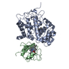
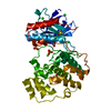
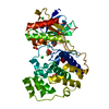
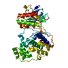
- PDB-3qvc: Crystal structure of histo-aspartic protease (HAP) zymogen from P... -
+ Open data

- Basic information
| Entry | Database: PDB / ID: 3qvc | ||||||
|---|---|---|---|---|---|---|---|
| Title | Crystal structure of histo-aspartic protease (HAP) zymogen from Plasmodium falciparum | ||||||
 Components Components | Histo-aspartic protease | ||||||
 Keywords Keywords |  HYDROLASE / Histo-aspartic protease / HAP / Plasmepsin / Aspartic protease / Malaria / Zymogen HYDROLASE / Histo-aspartic protease / HAP / Plasmepsin / Aspartic protease / Malaria / Zymogen | ||||||
| Function / homology |  Function and homology informationplasmepsin II / acquisition of nutrients from host / vacuolar lumen / food vacuole / aspartic-type endopeptidase activity / proteolysis / membrane Function and homology informationplasmepsin II / acquisition of nutrients from host / vacuolar lumen / food vacuole / aspartic-type endopeptidase activity / proteolysis / membraneSimilarity search - Function | ||||||
| Biological species |  Plasmodium falciparum (malaria parasite P. falciparum) Plasmodium falciparum (malaria parasite P. falciparum) | ||||||
| Method | X-RAY DIFFRACTION / MOLECULAR REPLACEMENT / Resolution: 2.1 Å | ||||||
 Authors Authors | Bhaumik, P. / Gustchina, A. / Wlodawer, A. | ||||||
 Citation Citation | Journal: Biochemistry / Year: 2011 Title: Structural insights into the activation and inhibition of histo-aspartic protease from Plasmodium falciparum. Authors: Bhaumik, P. / Xiao, H. / Hidaka, K. / Gustchina, A. / Kiso, Y. / Yada, R.Y. / Wlodawer, A. | ||||||
| History |
|
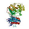
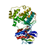
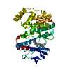
- Structure visualization
| Structure viewer | Molecule: MolmilJmol/JSmol |
|---|
- Downloads & links
-Download
| PDBx/mmCIF format | 3qvc.cif.gz | 164.1 KB | Display | PDBx/mmCIF format |
|---|---|---|---|---|
| PDB format | pdb3qvc.ent.gz | 135.6 KB | Display | PDB format |
| PDBx/mmJSON format | 3qvc.json.gz | Tree view | PDBx/mmJSON format | |
| Others |  Other downloads Other downloads |
-Validation report
| Arichive directory | https://data.pdbj.org/pub/pdb/validation_reports/qv/3qvcftp://data.pdbj.org/pub/pdb/validation_reports/qv/3qvc | HTTPS FTP |
|---|
-Related structure data


-Links
 PDBj
PDBj- Assembly
| Deposited unit | 
| ||||||||
|---|---|---|---|---|---|---|---|---|---|
| 1 |
| ||||||||
| 2 | 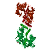
| ||||||||
| Unit cell |
|
-Components
| #1: Protein | Mass: 51754.973 Da / Num. of mol.: 1 Source method: isolated from a genetically manipulated source Source: (gene. exp.) Plasmodium falciparum (malaria parasite P. falciparum)Gene: hap / Plasmid: pET32B(+) / Production host:  Escherichia coli (E. coli) / Strain (production host): Rosetta-gami B (DE3) pLysS / References: UniProt: Q9Y006 Escherichia coli (E. coli) / Strain (production host): Rosetta-gami B (DE3) pLysS / References: UniProt: Q9Y006 | ||
|---|---|---|---|
| #2: Chemical | ChemComp-EDO / Ethylene glycol  Mass: 62.068 Da / Num. of mol.: 5 / Source method: obtained synthetically / Formula: C2H6O2 Mass: 62.068 Da / Num. of mol.: 5 / Source method: obtained synthetically / Formula: C2H6O2#3: Water | ChemComp-HOH / | Water Mass: 18.015 Da / Num. of mol.: 242 / Source method: isolated from a natural source / Formula: H2O Mass: 18.015 Da / Num. of mol.: 242 / Source method: isolated from a natural source / Formula: H2O |
-Experimental details
-Experiment
| Experiment | Method: X-RAY DIFFRACTION / Number of used crystals: 1 |
|---|
- Sample preparation
| Crystal | Density Matthews: 2.37 Å3/Da / Density % sol: 48.08 % |
|---|---|
| Crystal grow | Temperature: 293 K / Method: vapor diffusion, sitting drop / pH: 8.1 Details: 20% (w/v) PEG 3350, 0.2M Tri-potassium citrate, pH 8.1, VAPOR DIFFUSION, SITTING DROP, temperature 293K |
-Data collection
| Diffraction | Mean temperature: 100 K |
|---|---|
| Diffraction source | Source: ROTATING ANODE / Wavelength: 1.5418 Å |
| Detector | Type: MAR scanner 345 mm plate / Detector: IMAGE PLATE / Date: Jul 31, 2010 |
| Radiation | Protocol: SINGLE WAVELENGTH / Monochromatic (M) / Laue (L): M / Scattering type: x-ray |
| Radiation wavelength | Wavelength: 1.5418 Å / Relative weight: 1 |
| Reflection | Resolution: 1.9→40 Å / Num. all: 38417 / Num. obs: 37803 / % possible obs: 98.4 % / Redundancy: 5 % / Rmerge(I) obs: 0.102 / Net I/σ(I): 11.9 |
| Reflection shell | Resolution: 1.9→2 Å / Redundancy: 4.5 % / Rmerge(I) obs: 0.1461 / Mean I/σ(I) obs: 1.2 / % possible all: 97.1 |
- Processing
| Software |
| ||||||||||||||||||||||||||||||||||||||||||||||||||||||||||||||||||||||||||||||||||||||||||||||||||||||||||||||||||||||||||||||||||||||||||||||||||||||||||||||||||||||||||
|---|---|---|---|---|---|---|---|---|---|---|---|---|---|---|---|---|---|---|---|---|---|---|---|---|---|---|---|---|---|---|---|---|---|---|---|---|---|---|---|---|---|---|---|---|---|---|---|---|---|---|---|---|---|---|---|---|---|---|---|---|---|---|---|---|---|---|---|---|---|---|---|---|---|---|---|---|---|---|---|---|---|---|---|---|---|---|---|---|---|---|---|---|---|---|---|---|---|---|---|---|---|---|---|---|---|---|---|---|---|---|---|---|---|---|---|---|---|---|---|---|---|---|---|---|---|---|---|---|---|---|---|---|---|---|---|---|---|---|---|---|---|---|---|---|---|---|---|---|---|---|---|---|---|---|---|---|---|---|---|---|---|---|---|---|---|---|---|---|---|---|---|
| Refinement | Method to determine structure: MOLECULAR REPLACEMENT / Resolution: 2.1→36.37 Å / Cor.coef. Fo:Fc: 0.963 / Cor.coef. Fo:Fc free: 0.935 / SU B: 12.521 / SU ML: 0.144 / Cross valid method: THROUGHOUT / ESU R: 0.19 / ESU R Free: 0.178 / Stereochemistry target values: MAXIMUM LIKELIHOOD
| ||||||||||||||||||||||||||||||||||||||||||||||||||||||||||||||||||||||||||||||||||||||||||||||||||||||||||||||||||||||||||||||||||||||||||||||||||||||||||||||||||||||||||
| Solvent computation | Ion probe radii: 0.8 Å / Shrinkage radii: 0.8 Å / VDW probe radii: 1.2 Å / Solvent model: MASK | ||||||||||||||||||||||||||||||||||||||||||||||||||||||||||||||||||||||||||||||||||||||||||||||||||||||||||||||||||||||||||||||||||||||||||||||||||||||||||||||||||||||||||
| Displacement parameters | Biso mean: 40.768 Å2
| ||||||||||||||||||||||||||||||||||||||||||||||||||||||||||||||||||||||||||||||||||||||||||||||||||||||||||||||||||||||||||||||||||||||||||||||||||||||||||||||||||||||||||
| Refinement step | Cycle: LAST / Resolution: 2.1→36.37 Å
| ||||||||||||||||||||||||||||||||||||||||||||||||||||||||||||||||||||||||||||||||||||||||||||||||||||||||||||||||||||||||||||||||||||||||||||||||||||||||||||||||||||||||||
| Refine LS restraints |
| ||||||||||||||||||||||||||||||||||||||||||||||||||||||||||||||||||||||||||||||||||||||||||||||||||||||||||||||||||||||||||||||||||||||||||||||||||||||||||||||||||||||||||
| LS refinement shell | Resolution: 2.1→2.154 Å / Total num. of bins used: 20
| ||||||||||||||||||||||||||||||||||||||||||||||||||||||||||||||||||||||||||||||||||||||||||||||||||||||||||||||||||||||||||||||||||||||||||||||||||||||||||||||||||||||||||
| Refinement TLS params. | Method: refined / Refine-ID: X-RAY DIFFRACTION
| ||||||||||||||||||||||||||||||||||||||||||||||||||||||||||||||||||||||||||||||||||||||||||||||||||||||||||||||||||||||||||||||||||||||||||||||||||||||||||||||||||||||||||
| Refinement TLS group |
|
