 Movie
Movie Controller

[English] 日本語
 Yorodumi
- PDB-3qum: Crystal structure of human prostate specific antigen (PSA) in Fab... -
+ Open data

- Basic information
| Entry | Database: PDB / ID: 3qum | |||||||||
|---|---|---|---|---|---|---|---|---|---|---|
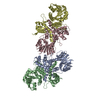
| Title | Crystal structure of human prostate specific antigen (PSA) in Fab sandwich with a high affinity and a PCa selective antibody | |||||||||
 Components Components |
| |||||||||
 Keywords Keywords |  IMMUNE SYSTEM / kallikrein fold / Prostate-specific antigen / serine protease / negative regulation of angiogenesis / natural post-transductional modification / N-linked and O-linked glycosylation IMMUNE SYSTEM / kallikrein fold / Prostate-specific antigen / serine protease / negative regulation of angiogenesis / natural post-transductional modification / N-linked and O-linked glycosylation | |||||||||
| Function / homology |  Function and homology informationsemenogelase / positive regulation of antibacterial peptide production / hydrolase activity, acting on carbon-nitrogen (but not peptide) bonds, in linear amides / protein metabolic process / serine-type peptidase activity / negative regulation of angiogenesis / secretory granule / Activated PKN1 stimulates transcription of AR (androgen receptor) regulated genes KLK2 and KLK3 / Regulation of Insulin-like Growth Factor (IGF) transport and uptake by Insulin-like Growth Factor Binding Proteins (IGFBPs) / endopeptidase activity ...semenogelase / positive regulation of antibacterial peptide production / hydrolase activity, acting on carbon-nitrogen (but not peptide) bonds, in linear amides / protein metabolic process / serine-type peptidase activity / negative regulation of angiogenesis / secretory granule / Activated PKN1 stimulates transcription of AR (androgen receptor) regulated genes KLK2 and KLK3 / Regulation of Insulin-like Growth Factor (IGF) transport and uptake by Insulin-like Growth Factor Binding Proteins (IGFBPs) / endopeptidase activity / serine-type endopeptidase activity / protein-containing complex / proteolysis / extracellular space / extracellular exosome / extracellular region / nucleus Function and homology informationsemenogelase / positive regulation of antibacterial peptide production / hydrolase activity, acting on carbon-nitrogen (but not peptide) bonds, in linear amides / protein metabolic process / serine-type peptidase activity / negative regulation of angiogenesis / secretory granule / Activated PKN1 stimulates transcription of AR (androgen receptor) regulated genes KLK2 and KLK3 / Regulation of Insulin-like Growth Factor (IGF) transport and uptake by Insulin-like Growth Factor Binding Proteins (IGFBPs) / endopeptidase activity ...semenogelase / positive regulation of antibacterial peptide production / hydrolase activity, acting on carbon-nitrogen (but not peptide) bonds, in linear amides / protein metabolic process / serine-type peptidase activity / negative regulation of angiogenesis / secretory granule / Activated PKN1 stimulates transcription of AR (androgen receptor) regulated genes KLK2 and KLK3 / Regulation of Insulin-like Growth Factor (IGF) transport and uptake by Insulin-like Growth Factor Binding Proteins (IGFBPs) / endopeptidase activity / serine-type endopeptidase activity / protein-containing complex / proteolysis / extracellular space / extracellular exosome / extracellular region / nucleusSimilarity search - Function | |||||||||
| Biological species |  Homo sapiens (human) Homo sapiens (human) Mus musculus (house mouse) Mus musculus (house mouse) | |||||||||
| Method | X-RAY DIFFRACTION / SYNCHROTRON / MOLECULAR REPLACEMENT / Resolution: 3.2 Å | |||||||||
 Authors Authors | Stura, E.A. / Muller, B.H. / Michel, S. / Ducancel, F. | |||||||||
 Citation Citation | Journal: J.Mol.Biol. / Year: 2011 Title: Crystal structure of human prostate-specific antigen in a sandwich antibody complex. Authors: Stura, E.A. / Muller, B.H. / Bossus, M. / Michel, S. / Jolivet-Reynaud, C. / Ducancel, F. #1: Journal: J.Mol.Biol. / Year: 2011 Title: In Vitro Affinity Maturation of an Anti-PSA Antibody for Prostate Cancer Diagnostic Assay. Authors: Muller, B.H. / Savatier, A. / L'hostis, G. / Costa, N. / Bossus, M. / Michel, S. / Ott, C. / Becquart, L. / Ruffion, A. / Stura, E.A. / Ducancel, F. | |||||||||
| History |
|
- Structure visualization
| Structure viewer | Molecule: MolmilJmol/JSmol |
|---|
- Downloads & links
-Download
| PDBx/mmCIF format | 3qum.cif.gz | 451 KB | Display | PDBx/mmCIF format |
|---|---|---|---|---|
| PDB format | pdb3qum.ent.gz | 364.4 KB | Display | PDB format |
| PDBx/mmJSON format | 3qum.json.gz | Tree view | PDBx/mmJSON format | |
| Others |  Other downloads Other downloads |
-Validation report
| Arichive directory | https://data.pdbj.org/pub/pdb/validation_reports/qu/3qumftp://data.pdbj.org/pub/pdb/validation_reports/qu/3qum | HTTPS FTP |
|---|
-Related structure data
| Related structure data |  2zchS S: Starting model for refinement |
|---|---|
| Similar structure data |


-Links
 PDBj
PDBj

- Assembly
| Deposited unit | 
| ||||||||
|---|---|---|---|---|---|---|---|---|---|
| 1 | 
| ||||||||
| 2 | 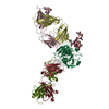
| ||||||||
| Unit cell |
|
-Components
-Antibody , 4 types, 8 molecules LMHKACBD
| #2: Antibody | Mass: 24271.965 Da / Num. of mol.: 2 / Source method: isolated from a natural source / Source: (natural) Mus musculus (house mouse)#3: Antibody | Mass: 23551.430 Da / Num. of mol.: 2 / Source method: isolated from a natural source / Source: (natural) Mus musculus (house mouse)#4: Antibody | Mass: 24189.725 Da / Num. of mol.: 2 / Source method: isolated from a natural source / Source: (natural) Mus musculus (house mouse)#5: Antibody | Mass: 23565.393 Da / Num. of mol.: 2 / Source method: isolated from a natural source / Source: (natural) Mus musculus (house mouse) |
|---|
-Protein / Non-polymers , 2 types, 93 molecules PQ
| #1: Protein | / PSA / Gamma-seminoprotein / Seminin / Kallikrein-3 / P-30 antigen / Semenogelase Mass: 26122.025 Da / Num. of mol.: 2 / Source method: isolated from a natural source / Source: (natural) Homo sapiens (human) / References: UniProt: P07288, semenogelase#9: Water | ChemComp-HOH / | WaterMass: 18.015 Da / Num. of mol.: 91 / Source method: isolated from a natural source / Formula: H2O |
|---|
-Sugars , 3 types, 3 molecules
| #6: Polysaccharide | N-acetyl-alpha-neuraminic acid-(2-3)-beta-D-galactopyranose-(1-4)-2-acetamido-2-deoxy-beta-D- ...N-acetyl-alpha-neuraminic acid-(2-3)-beta-D-galactopyranose-(1-4)-2-acetamido-2-deoxy-beta-D-glucopyranose-(1-4)-alpha-D-mannopyranose-(1-3)-[N-acetyl-alpha-neuraminic acid-(2-3)-beta-D-galactopyranose-(1-4)-2-acetamido-2-deoxy-beta-D-glucopyranose-(1-2)-[N-acetyl-alpha-neuraminic acid-(2-3)-beta-D-galactopyranose-(1-4)-2-acetamido-2-deoxy-beta-D-glucopyranose-(1-6)]alpha-D-mannopyranose-(1-6)]alpha-D-mannopyranose-(1-4)-2-acetamido-2-deoxy-beta-D-glucopyranose-(1-4)-[alpha-L-fucopyranose-(1-6)]2-acetamido-2-deoxy-beta-D-glucopyranose / Mass: 3026.729 Da / Num. of mol.: 1 Source method: isolated from a genetically manipulated source |
|---|---|
| #7: Polysaccharide | beta-D-galactopyranose-(1-3)-2-acetamido-2-deoxy-alpha-D-galactopyranose / Thomsen-Friedenreich antigen / Thomsen–Friedenreich antigen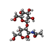  , Oligosaccharide / Class: Antigen / Mass: 383.349 Da / Num. of mol.: 1 , Oligosaccharide / Class: Antigen / Mass: 383.349 Da / Num. of mol.: 1Source method: isolated from a genetically manipulated source Details: oligosaccharide / References: Thomsen-Friedenreich antigen |
| #8: Polysaccharide | N-acetyl-alpha-neuraminic acid-(2-3)-beta-D-galactopyranose-(1-4)-2-acetamido-2-deoxy-beta-D- ...N-acetyl-alpha-neuraminic acid-(2-3)-beta-D-galactopyranose-(1-4)-2-acetamido-2-deoxy-beta-D-glucopyranose-(1-6)-alpha-D-mannopyranose-(1-3)-[N-acetyl-alpha-neuraminic acid-(2-3)-beta-D-galactopyranose-(1-4)-2-acetamido-2-deoxy-beta-D-glucopyranose-(1-2)-[N-acetyl-alpha-neuraminic acid-(2-3)-beta-D-galactopyranose-(1-4)-2-acetamido-2-deoxy-beta-D-glucopyranose-(1-6)]alpha-D-mannopyranose-(1-6)]alpha-D-mannopyranose-(1-4)-2-acetamido-2-deoxy-beta-D-glucopyranose-(1-4)-[alpha-L-fucopyranose-(1-6)]2-acetamido-2-deoxy-beta-D-glucopyranose / Mass: 3026.729 Da / Num. of mol.: 1 Source method: isolated from a genetically manipulated source |
-Experimental details
-Experiment
| Experiment | Method: X-RAY DIFFRACTION / Number of used crystals: 1 |
|---|
- Sample preparation
| Crystal | Density Matthews: 3.04 Å3/Da / Density % sol: 59.54 % |
|---|---|
| Crystal grow | Temperature: 295 K / Method: vapor diffusion, sitting drop / pH: 6.5 Details: Protein solution: 20 ul 5D3D11 at 10.7 mg/ml and 10 ul PSA at 5 mg/ml + 31 ul 5D5A5 at 6.7mg/ml in PBS. against a precipitant of 10% PEG 4,000, 20% ethylene glycol, 100 mM Na,K phosphate. ...Details: Protein solution: 20 ul 5D3D11 at 10.7 mg/ml and 10 ul PSA at 5 mg/ml + 31 ul 5D5A5 at 6.7mg/ml in PBS. against a precipitant of 10% PEG 4,000, 20% ethylene glycol, 100 mM Na,K phosphate. Drops consisted of 1 ul protein + precipitant equilibrated by vapor diffusion. Streak seeding and macro seeding were used, pH 6.5, VAPOR DIFFUSION, SITTING DROP, temperature 295K |
-Data collection
| Diffraction | Mean temperature: 100 K | |||||||||||||||||||||||||||||||||||||||||||||||||||||||||||||||||||||||||||||
|---|---|---|---|---|---|---|---|---|---|---|---|---|---|---|---|---|---|---|---|---|---|---|---|---|---|---|---|---|---|---|---|---|---|---|---|---|---|---|---|---|---|---|---|---|---|---|---|---|---|---|---|---|---|---|---|---|---|---|---|---|---|---|---|---|---|---|---|---|---|---|---|---|---|---|---|---|---|---|
| Diffraction source | Source: SYNCHROTRON / Site: ESRF  / Beamline: ID23-2 / Wavelength: 0.8726 Å / Beamline: ID23-2 / Wavelength: 0.8726 Å | |||||||||||||||||||||||||||||||||||||||||||||||||||||||||||||||||||||||||||||
| Detector | Type: MARMOSAIC 225 mm CCD / Detector: CCD / Date: Mar 17, 2007 Details: Pt coated mirrors in a Kirkpatrick-Baez (KB) geometry | |||||||||||||||||||||||||||||||||||||||||||||||||||||||||||||||||||||||||||||
| Radiation | Monochromator: diffracting Si (111) monochromator / Protocol: SINGLE WAVELENGTH / Monochromatic (M) / Laue (L): M / Scattering type: x-ray | |||||||||||||||||||||||||||||||||||||||||||||||||||||||||||||||||||||||||||||
| Radiation wavelength | Wavelength: 0.8726 Å / Relative weight: 1 | |||||||||||||||||||||||||||||||||||||||||||||||||||||||||||||||||||||||||||||
| Reflection | Resolution: 3.2→81.65 Å / Num. all: 47764 / Num. obs: 47573 / % possible obs: 99.6 % / Observed criterion σ(F): 0 / Observed criterion σ(I): -3 / Redundancy: 2.5 % / Biso Wilson estimate: 83.924 Å2 / Rmerge(I) obs: 0.145 / Rsym value: 0.145 / Net I/σ(I): 8.4 | |||||||||||||||||||||||||||||||||||||||||||||||||||||||||||||||||||||||||||||
| Reflection shell | Diffraction-ID: 1
|
- Processing
| Software |
| ||||||||||||||||||||||||||||||||||||||||||||||||||||||||||||||||||||||||||||||||||||||||||||||||||||||||||||||||||||||||||||||
|---|---|---|---|---|---|---|---|---|---|---|---|---|---|---|---|---|---|---|---|---|---|---|---|---|---|---|---|---|---|---|---|---|---|---|---|---|---|---|---|---|---|---|---|---|---|---|---|---|---|---|---|---|---|---|---|---|---|---|---|---|---|---|---|---|---|---|---|---|---|---|---|---|---|---|---|---|---|---|---|---|---|---|---|---|---|---|---|---|---|---|---|---|---|---|---|---|---|---|---|---|---|---|---|---|---|---|---|---|---|---|---|---|---|---|---|---|---|---|---|---|---|---|---|---|---|---|---|
| Refinement | Method to determine structure: MOLECULAR REPLACEMENT Starting model: PDB entry 2ZCH, using individual PSA and Fab as independent search models. Resolution: 3.2→58.965 Å / SU ML: 0.48 / Isotropic thermal model: Isotropic / Cross valid method: THROUGHOUT / σ(F): 1.36 / σ(I): -3 / Stereochemistry target values: ML
| ||||||||||||||||||||||||||||||||||||||||||||||||||||||||||||||||||||||||||||||||||||||||||||||||||||||||||||||||||||||||||||||
| Solvent computation | Shrinkage radii: 0.83 Å / VDW probe radii: 1.1 Å / Solvent model: FLAT BULK SOLVENT MODEL / Bsol: 68.872 Å2 / ksol: 0.304 e/Å3 | ||||||||||||||||||||||||||||||||||||||||||||||||||||||||||||||||||||||||||||||||||||||||||||||||||||||||||||||||||||||||||||||
| Displacement parameters | Biso mean: 73.269 Å2
| ||||||||||||||||||||||||||||||||||||||||||||||||||||||||||||||||||||||||||||||||||||||||||||||||||||||||||||||||||||||||||||||
| Refinement step | Cycle: LAST / Resolution: 3.2→58.965 Å
| ||||||||||||||||||||||||||||||||||||||||||||||||||||||||||||||||||||||||||||||||||||||||||||||||||||||||||||||||||||||||||||||
| Refine LS restraints |
| ||||||||||||||||||||||||||||||||||||||||||||||||||||||||||||||||||||||||||||||||||||||||||||||||||||||||||||||||||||||||||||||
| LS refinement shell |
|
