 Movie
Movie Controller

[English] 日本語
 Yorodumi
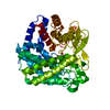
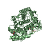
- PDB-3qry: Analysis of a new family of widely distributed metal-independent ... -
+ Open data

- Basic information
| Entry | Database: PDB / ID: 3qry | ||||||
|---|---|---|---|---|---|---|---|
| Title | Analysis of a new family of widely distributed metal-independent alpha mannosidases provides unique insight into the processing of N-linked glycans, Streptococcus pneumoniae SP_2144 1-deoxymannojirimycin complex | ||||||
 Components Components | Putative uncharacterized protein | ||||||
 Keywords Keywords |  HYDROLASE / ALPHA-ALPHA SIX FOLD / GLYCOSIDE HYDROLASE / MANNOSIDASE / 1-DEOXYMANNOJIRIMYCIN HYDROLASE / ALPHA-ALPHA SIX FOLD / GLYCOSIDE HYDROLASE / MANNOSIDASE / 1-DEOXYMANNOJIRIMYCIN | ||||||
| Function / homology |  Function and homology information Function and homology informationcarbohydrate catabolic process / catalytic activity / carbohydrate metabolic processSimilarity search - Function | ||||||
| Biological species |   Streptococcus pneumoniae (bacteria) Streptococcus pneumoniae (bacteria) | ||||||
| Method | X-RAY DIFFRACTION / SYNCHROTRON / MOLECULAR REPLACEMENT / Resolution: 1.75 Å | ||||||
 Authors Authors | Gregg, K.J. / Zandberg, W.F. / Hehemann, J.-H. / Whitworth, G.E. / Deng, L.E. / Vocadlo, D.J. / Boraston, A.B. | ||||||
 Citation Citation | Journal: J.Biol.Chem. / Year: 2011 Title: Analysis of a New Family of Widely Distributed Metal-independent {alpha}-Mannosidases Provides Unique Insight into the Processing of N-Linked Glycans. Authors: Gregg, K.J. / Zandberg, W.F. / Hehemann, J.H. / Whitworth, G.E. / Deng, L. / Vocadlo, D.J. / Boraston, A.B. | ||||||
| History |
|
- Structure visualization
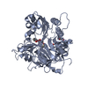
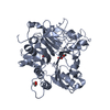
| Structure viewer | Molecule: MolmilJmol/JSmol |
|---|
- Downloads & links
-Download
| PDBx/mmCIF format | 3qry.cif.gz | 213.6 KB | Display | PDBx/mmCIF format |
|---|---|---|---|---|
| PDB format | pdb3qry.ent.gz | 169.7 KB | Display | PDB format |
| PDBx/mmJSON format | 3qry.json.gz | Tree view | PDBx/mmJSON format | |
| Others |  Other downloads Other downloads |
-Validation report
| Arichive directory | https://data.pdbj.org/pub/pdb/validation_reports/qr/3qryftp://data.pdbj.org/pub/pdb/validation_reports/qr/3qry | HTTPS FTP |
|---|
-Related structure data


-Links
 PDBj
PDBj
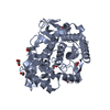
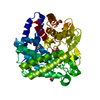
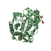
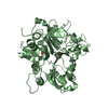
- Assembly
| Deposited unit | 
| ||||||||
|---|---|---|---|---|---|---|---|---|---|
| 1 | 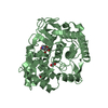
| ||||||||
| 2 | 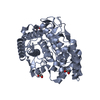
| ||||||||
| Unit cell |
|
-Components
| #1: Protein | Mass: 49105.828 Da / Num. of mol.: 2 Source method: isolated from a genetically manipulated source Source: (gene. exp.) Streptococcus pneumoniae (bacteria) / Gene: SP_2144 / Production host: Escherichia coli (E. coli) / Strain (production host): Bl21 DE3 STAR / References: UniProt: Q97NA8, UniProt: A0A0H2URZ6*PLUS#2: Chemical | 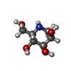  Mass: 163.172 Da / Num. of mol.: 2 / Source method: obtained synthetically / Formula: C6H13NO4 Mass: 163.172 Da / Num. of mol.: 2 / Source method: obtained synthetically / Formula: C6H13NO4#3: Chemical | ChemComp-EDO / Ethylene glycol  Mass: 62.068 Da / Num. of mol.: 6 / Source method: obtained synthetically / Formula: C2H6O2 Mass: 62.068 Da / Num. of mol.: 6 / Source method: obtained synthetically / Formula: C2H6O2#4: Water | ChemComp-HOH / | Water Mass: 18.015 Da / Num. of mol.: 1388 / Source method: isolated from a natural source / Formula: H2O Mass: 18.015 Da / Num. of mol.: 1388 / Source method: isolated from a natural source / Formula: H2O |
|---|
-Experimental details
-Experiment
| Experiment | Method: X-RAY DIFFRACTION / Number of used crystals: 1 |
|---|
- Sample preparation
| Crystal | Density Matthews: 2.48 Å3/Da / Density % sol: 50.38 % |
|---|---|
| Crystal grow | Method: vapor diffusion, hanging drop / pH: 8 Details: 0.1 M Hepes, 1.2 M LiSO4, pH 8.0, VAPOR DIFFUSION, HANGING DROP |
-Data collection
| Diffraction | Mean temperature: 117 K |
|---|---|
| Diffraction source | Source: SYNCHROTRON / Site: SSRL  / Beamline: BL9-2 / Wavelength: 0.97946 Å / Beamline: BL9-2 / Wavelength: 0.97946 Å |
| Detector | Type: MARMOSAIC 325 mm CCD / Detector: CCD / Date: Jan 1, 2010 |
| Radiation | Monochromator: Double crystal monochromator / Protocol: SINGLE WAVELENGTH / Monochromatic (M) / Laue (L): M / Scattering type: x-ray |
| Radiation wavelength | Wavelength: 0.97946 Å / Relative weight: 1 |
| Reflection | Resolution: 1.75→158.8 Å / Num. all: 88818 / Num. obs: 88818 / % possible obs: 99 % / Observed criterion σ(F): 2 / Observed criterion σ(I): 2 |
| Reflection shell | Resolution: 1.75→1.84 Å / % possible all: 2 |
- Processing
| Software |
| |||||||||||||||||||||||||||||||||||||||||||||||||||||||||||||||||
|---|---|---|---|---|---|---|---|---|---|---|---|---|---|---|---|---|---|---|---|---|---|---|---|---|---|---|---|---|---|---|---|---|---|---|---|---|---|---|---|---|---|---|---|---|---|---|---|---|---|---|---|---|---|---|---|---|---|---|---|---|---|---|---|---|---|---|
| Refinement | Method to determine structure: MOLECULAR REPLACEMENT / Resolution: 1.75→20 Å / Cor.coef. Fo:Fc: 0.95 / Cor.coef. Fo:Fc free: 0.916 / SU B: 2.226 / SU ML: 0.073 / Cross valid method: THROUGHOUT / σ(F): 2 / ESU R Free: 0.12 / Stereochemistry target values: MAXIMUM LIKELIHOOD / Details: HYDROGENS HAVE BEEN ADDED IN THE RIDING POSITIONS
| |||||||||||||||||||||||||||||||||||||||||||||||||||||||||||||||||
| Solvent computation | Ion probe radii: 0.8 Å / Shrinkage radii: 0.8 Å / VDW probe radii: 1.4 Å / Solvent model: MASK | |||||||||||||||||||||||||||||||||||||||||||||||||||||||||||||||||
| Displacement parameters | Biso mean: 13.151 Å2
| |||||||||||||||||||||||||||||||||||||||||||||||||||||||||||||||||
| Refinement step | Cycle: LAST / Resolution: 1.75→20 Å
| |||||||||||||||||||||||||||||||||||||||||||||||||||||||||||||||||
| Refine LS restraints |
| |||||||||||||||||||||||||||||||||||||||||||||||||||||||||||||||||
| LS refinement shell | Resolution: 1.75→1.795 Å / Total num. of bins used: 20
|
