 Movie
Movie Controller

[English] 日本語
 Yorodumi
- PDB-3qqn: The retinal specific CD147 Ig0 domain: from molecular structure t... -
+ Open data

- Basic information
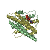
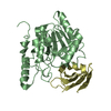
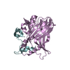
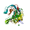
| Entry | Database: PDB / ID: 3qqn | ||||||
|---|---|---|---|---|---|---|---|
| Title | The retinal specific CD147 Ig0 domain: from molecular structure to biological activity | ||||||
 Components Components | Basigin | ||||||
 Keywords Keywords | CELL ADHESION / CD147 / EMMPRIN / Immunoglobulin-like domain / beta sheet / Structural Genomics / Berkeley Structural Genomics Center / BSGC | ||||||
| Function / homology |  Function and homology information Function and homology informationDefective SLC16A1 causes symptomatic deficiency in lactate transport (SDLT) / Proton-coupled monocarboxylate transport / positive regulation of matrix metallopeptidase secretion / acrosomal membrane / response to mercury ion / neural retina development / endothelial tube morphogenesis / Pyruvate metabolism / photoreceptor cell maintenance / Basigin interactions ...Defective SLC16A1 causes symptomatic deficiency in lactate transport (SDLT) / Proton-coupled monocarboxylate transport / positive regulation of matrix metallopeptidase secretion / acrosomal membrane / response to mercury ion / neural retina development / endothelial tube morphogenesis / Pyruvate metabolism / photoreceptor cell maintenance / Basigin interactions / Aspirin ADME / odontogenesis of dentin-containing tooth / D-mannose binding / decidualization / photoreceptor outer segment / positive regulation of vascular endothelial growth factor production / Integrin cell surface interactions / embryo implantation / response to cAMP / photoreceptor inner segment / Degradation of the extracellular matrix / neutrophil chemotaxis / positive regulation of endothelial cell migration / protein localization to plasma membrane / sarcolemma / response to peptide hormone / positive regulation of interleukin-6 production / melanosome / virus receptor activity / signaling receptor activity / basolateral plasma membrane / angiogenesis / positive regulation of viral entry into host cell / cell surface receptor signaling pathway / endosome / cadherin binding / Golgi membrane / focal adhesion / intracellular membrane-bounded organelle / endoplasmic reticulum membrane / mitochondrion / extracellular exosome / membrane / plasma membraneSimilarity search - Function | ||||||
| Biological species |  Homo sapiens (human) Homo sapiens (human) | ||||||
| Method | X-RAY DIFFRACTION / SYNCHROTRON / Resolution: 2.309 Å | ||||||
 Authors Authors | Redzic, J.S. / Armstrong, G.S. / Isern, N.G. / Kieft, J.S. / Eisenmesser, E.Z. / Berkeley Structural Genomics Center (BSGC) | ||||||
 Citation Citation | Journal: J.Mol.Biol. / Year: 2011 Title: The Retinal Specific CD147 Ig0 Domain: From Molecular Structure to Biological Activity. Authors: Redzic, J.S. / Armstrong, G.S. / Isern, N.G. / Jones, D.N. / Kieft, J.S. / Eisenmesser, E.Z. | ||||||
| History |
|
- Structure visualization
| Structure viewer | Molecule: MolmilJmol/JSmol |
|---|
- Downloads & links
-Download
| PDBx/mmCIF format | 3qqn.cif.gz | 59.9 KB | Display | PDBx/mmCIF format |
|---|---|---|---|---|
| PDB format | pdb3qqn.ent.gz | 48.3 KB | Display | PDB format |
| PDBx/mmJSON format | 3qqn.json.gz | Tree view | PDBx/mmJSON format | |
| Others |  Other downloads Other downloads |
-Validation report
| Arichive directory | https://data.pdbj.org/pub/pdb/validation_reports/qq/3qqnftp://data.pdbj.org/pub/pdb/validation_reports/qq/3qqn | HTTPS FTP |
|---|
-Related structure data


-Links
 PDBj
PDBj


- Assembly
| Deposited unit | 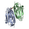
| ||||||||||||||||||
|---|---|---|---|---|---|---|---|---|---|---|---|---|---|---|---|---|---|---|---|
| 1 |
| ||||||||||||||||||
| Unit cell |
| ||||||||||||||||||
| Components on special symmetry positions |
| ||||||||||||||||||
| Noncrystallographic symmetry (NCS) | NCS domain:
NCS domain segments:
|
-Components
| #1: Antibody | / 5F7 / Collagenase stimulatory factor / Extracellular matrix metalloproteinase inducer / EMMPRIN / ...5F7 / Collagenase stimulatory factor / Extracellular matrix metalloproteinase inducer / EMMPRIN / Leukocyte activation antigen M6 / OK blood group antigen / Tumor cell-derived collagenase stimulatory factor / TCSF Mass: 15469.710 Da / Num. of mol.: 2 / Fragment: UNP residues 23-138 / Mutation: C46M Source method: isolated from a genetically manipulated source Source: (gene. exp.) Homo sapiens (human) / Gene: BSG, UNQ6505/PRO21383 / Production host:  Escherichia coli (E. coli) / References: UniProt: P35613 Escherichia coli (E. coli) / References: UniProt: P35613#2: Water | ChemComp-HOH / | Water Mass: 18.015 Da / Num. of mol.: 158 / Source method: isolated from a natural source / Formula: H2O Mass: 18.015 Da / Num. of mol.: 158 / Source method: isolated from a natural source / Formula: H2OSequence details | AS PER AUTHORS ILE IS THE CORRECT RESIDUE AT POSITION 45 | |
|---|
-Experimental details
-Experiment
| Experiment | Method: X-RAY DIFFRACTION / Number of used crystals: 1 |
|---|
- Sample preparation
| Crystal | Density Matthews: 2.36 Å3/Da / Density % sol: 47.9 % |
|---|---|
| Crystal grow | Temperature: 277 K / Method: vapor diffusion, sitting drop / pH: 7 Details: 0.2M Ammonium Sulfate, 20% PEG 3350, 0.1M spermidine tetrachloride, pH 7, VAPOR DIFFUSION, SITTING DROP, temperature 277K |
-Data collection
| Diffraction | Mean temperature: 100 K | ||||||||||||
|---|---|---|---|---|---|---|---|---|---|---|---|---|---|
| Diffraction source | Source: SYNCHROTRON / Site: ALS  / Beamline: 4.2.2 / Wavelength: 1.02159,1.02195,1.03726 / Beamline: 4.2.2 / Wavelength: 1.02159,1.02195,1.03726 | ||||||||||||
| Detector | Type: NOIR-1 / Detector: CCD / Date: Oct 28, 2009 | ||||||||||||
| Radiation | Monochromator: Rosenbaum-Rock double Si111 crystal / Protocol: MAD / Monochromatic (M) / Laue (L): M / Scattering type: x-ray | ||||||||||||
| Radiation wavelength |
| ||||||||||||
| Reflection | Resolution: 2.309→42.42 Å / Num. obs: 14020 / % possible obs: 100 % / Observed criterion σ(F): 0 / Observed criterion σ(I): 0 |
- Processing
| Software |
| |||||||||||||||||||||||||||||||||||||||||||||||||||||||||||||||||||||||||||||
|---|---|---|---|---|---|---|---|---|---|---|---|---|---|---|---|---|---|---|---|---|---|---|---|---|---|---|---|---|---|---|---|---|---|---|---|---|---|---|---|---|---|---|---|---|---|---|---|---|---|---|---|---|---|---|---|---|---|---|---|---|---|---|---|---|---|---|---|---|---|---|---|---|---|---|---|---|---|---|
| Refinement | Resolution: 2.309→42.42 Å / SU ML: 0.29 / σ(F): 0 / Phase error: 25.77 / Stereochemistry target values: MLHL
| |||||||||||||||||||||||||||||||||||||||||||||||||||||||||||||||||||||||||||||
| Solvent computation | Shrinkage radii: 0.61 Å / VDW probe radii: 0.9 Å / Solvent model: FLAT BULK SOLVENT MODEL / Bsol: 40 Å2 / ksol: 0.302 e/Å3 | |||||||||||||||||||||||||||||||||||||||||||||||||||||||||||||||||||||||||||||
| Displacement parameters |
| |||||||||||||||||||||||||||||||||||||||||||||||||||||||||||||||||||||||||||||
| Refinement step | Cycle: LAST / Resolution: 2.309→42.42 Å
| |||||||||||||||||||||||||||||||||||||||||||||||||||||||||||||||||||||||||||||
| Refine LS restraints |
| |||||||||||||||||||||||||||||||||||||||||||||||||||||||||||||||||||||||||||||
| Refine LS restraints NCS |
| |||||||||||||||||||||||||||||||||||||||||||||||||||||||||||||||||||||||||||||
| LS refinement shell |
|
