 Movie
Movie Controller

[English] 日本語
 Yorodumi
- PDB-3qed: The structure and function of an arabinan-specific alpha-1,2-arab... -
+ Open data

- Basic information
| Entry | Database: PDB / ID: 3qed | ||||||
|---|---|---|---|---|---|---|---|
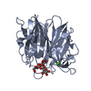
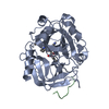
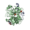
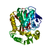
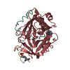
| Title | The structure and function of an arabinan-specific alpha-1,2-arabinofuranosidase identified from screening the activities of bacterial GH43 glycoside hydrolases | ||||||
 Components Components | Beta-xylosidase/alpha-L-arabinfuranosidase, gly43N | ||||||
 Keywords Keywords |  HYDROLASE / 5-bladed beta propeller HYDROLASE / 5-bladed beta propeller | ||||||
| Function / homology |  Function and homology informationHydrolases; Glycosylases; Glycosidases, i.e. enzymes that hydrolyse O- and S-glycosyl compounds / xylan catabolic process / hydrolase activity, hydrolyzing O-glycosyl compounds / metal ion binding Function and homology informationHydrolases; Glycosylases; Glycosidases, i.e. enzymes that hydrolyse O- and S-glycosyl compounds / xylan catabolic process / hydrolase activity, hydrolyzing O-glycosyl compounds / metal ion bindingSimilarity search - Function | ||||||
| Biological species |  Cellvibrio japonicus (bacteria) Cellvibrio japonicus (bacteria) | ||||||
| Method | X-RAY DIFFRACTION / SYNCHROTRON / SAD / Resolution: 2.99 Å | ||||||
 Authors Authors | Cartmell, A. / Mckee, L.S. / Pena, M. / Larsbrink, J. / Brumer, H. / Lewis, R.J. / Viks-Nielsen, A. / Gilbert, H.J. / Marles-Wright, J. | ||||||
 Citation Citation | Journal: J.Biol.Chem. / Year: 2011 Title: The Structure and Function of an Arabinan-specific {alpha}-1,2-Arabinofuranosidase Identified from Screening the Activities of Bacterial GH43 Glycoside Hydrolases. Authors: Cartmell, A. / McKee, L.S. / Pena, M.J. / Larsbrink, J. / Brumer, H. / Kaneko, S. / Ichinose, H. / Lewis, R.J. / Vikso-Nielsen, A. / Gilbert, H.J. / Marles-Wright, J. | ||||||
| History |
|
- Structure visualization
| Structure viewer | Molecule: MolmilJmol/JSmol |
|---|
- Downloads & links
-Download
| PDBx/mmCIF format | 3qed.cif.gz | 247.1 KB | Display | PDBx/mmCIF format |
|---|---|---|---|---|
| PDB format | pdb3qed.ent.gz | 210.7 KB | Display | PDB format |
| PDBx/mmJSON format | 3qed.json.gz | Tree view | PDBx/mmJSON format | |
| Others |  Other downloads Other downloads |
-Validation report
| Arichive directory | https://data.pdbj.org/pub/pdb/validation_reports/qe/3qedftp://data.pdbj.org/pub/pdb/validation_reports/qe/3qed | HTTPS FTP |
|---|
-Related structure data


-Links
 PDBj
PDBj- Assembly
| Deposited unit | 
| ||||||||
|---|---|---|---|---|---|---|---|---|---|
| 1 | 
| ||||||||
| 2 | 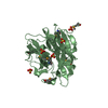
| ||||||||
| 3 | 
| ||||||||
| 4 | 
| ||||||||
| Unit cell |
|
-Components
| #1: Protein | Mass: 35686.719 Da / Num. of mol.: 4 / Fragment: UNP residues 28-334 Source method: isolated from a genetically manipulated source Source: (gene. exp.) Cellvibrio japonicus (bacteria) / Strain: Ueda107 / Gene: gly43N, CJA_3018 / Production host: Escherichia coli (E. coli) / Strain (production host): B834 (DE3)References: UniProt: B3PD60, Hydrolases; Glycosylases; Glycosidases, i.e. enzymes that hydrolyse O- and S-glycosyl compounds#2: Chemical | ChemComp-SO4 / Sulfate  Mass: 96.063 Da / Num. of mol.: 44 / Source method: obtained synthetically / Formula: SO4 Mass: 96.063 Da / Num. of mol.: 44 / Source method: obtained synthetically / Formula: SO4#3: Chemical | ChemComp-CA /   Mass: 40.078 Da / Num. of mol.: 4 / Source method: obtained synthetically / Formula: Ca Mass: 40.078 Da / Num. of mol.: 4 / Source method: obtained synthetically / Formula: Ca#4: Chemical | ChemComp-TAM / | Tris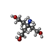  Mass: 163.215 Da / Num. of mol.: 1 / Source method: obtained synthetically / Formula: C7H17NO3 / Comment: pH buffer*YM Mass: 163.215 Da / Num. of mol.: 1 / Source method: obtained synthetically / Formula: C7H17NO3 / Comment: pH buffer*YM |
|---|
-Experimental details
-Experiment
| Experiment | Method: X-RAY DIFFRACTION / Number of used crystals: 1 |
|---|
- Sample preparation
| Crystal | Density Matthews: 4.3 Å3/Da / Density % sol: 71.38 % |
|---|---|
| Crystal grow | Temperature: 293 K / Method: vapor diffusion, hanging drop / pH: 7.5 Details: 1.5 M ammonium sulfate, 0.1 M Tris, pH 7.5, VAPOR DIFFUSION, HANGING DROP, temperature 293K |
-Data collection
| Diffraction | Mean temperature: 100 K |
|---|---|
| Diffraction source | Source: SYNCHROTRON / Site: Diamond  / Beamline: I02 / Wavelength: 0.98 / Beamline: I02 / Wavelength: 0.98 |
| Detector | Type: ADSC QUANTUM 315 / Detector: CCD / Date: Jul 1, 2008 |
| Radiation | Protocol: SINGLE WAVELENGTH / Monochromatic (M) / Laue (L): M / Scattering type: x-ray |
| Radiation wavelength | Wavelength: 0.98 Å / Relative weight: 1 |
| Reflection | Resolution: 2.99→72.17 Å / Num. obs: 50829 / % possible obs: 100 % / Observed criterion σ(F): 1 / Observed criterion σ(I): 2 / Redundancy: 14.3 % / Rmerge(I) obs: 0.215 / Net I/σ(I): 14.9 |
| Reflection shell | Resolution: 2.99→3.15 Å / Redundancy: 14.7 % / Rmerge(I) obs: 0.569 / Mean I/σ(I) obs: 5.2 / % possible all: 100 |
- Processing
| Software |
| ||||||||||||||||||||||||||||||||||||||||||||||||||||||||||||||||||||||||||||||||||||||||||||||||||||||||||||||||||||||||||||||||||||||||||||||||||||||||||||||||||||||||||
|---|---|---|---|---|---|---|---|---|---|---|---|---|---|---|---|---|---|---|---|---|---|---|---|---|---|---|---|---|---|---|---|---|---|---|---|---|---|---|---|---|---|---|---|---|---|---|---|---|---|---|---|---|---|---|---|---|---|---|---|---|---|---|---|---|---|---|---|---|---|---|---|---|---|---|---|---|---|---|---|---|---|---|---|---|---|---|---|---|---|---|---|---|---|---|---|---|---|---|---|---|---|---|---|---|---|---|---|---|---|---|---|---|---|---|---|---|---|---|---|---|---|---|---|---|---|---|---|---|---|---|---|---|---|---|---|---|---|---|---|---|---|---|---|---|---|---|---|---|---|---|---|---|---|---|---|---|---|---|---|---|---|---|---|---|---|---|---|---|---|---|---|
| Refinement | Method to determine structure: SAD / Resolution: 2.99→72.17 Å / Cor.coef. Fo:Fc: 0.907 / Cor.coef. Fo:Fc free: 0.876 / SU B: 12.463 / SU ML: 0.231 / Cross valid method: THROUGHOUT / σ(F): 1 / ESU R: 0.646 / ESU R Free: 0.325 / Stereochemistry target values: MAXIMUM LIKELIHOOD Details: HYDROGENS HAVE BEEN ADDED IN THE RIDING POSITIONS U VALUES : REFINED INDIVIDUALLY
| ||||||||||||||||||||||||||||||||||||||||||||||||||||||||||||||||||||||||||||||||||||||||||||||||||||||||||||||||||||||||||||||||||||||||||||||||||||||||||||||||||||||||||
| Solvent computation | Ion probe radii: 0.8 Å / Shrinkage radii: 0.8 Å / VDW probe radii: 1.4 Å / Solvent model: MASK | ||||||||||||||||||||||||||||||||||||||||||||||||||||||||||||||||||||||||||||||||||||||||||||||||||||||||||||||||||||||||||||||||||||||||||||||||||||||||||||||||||||||||||
| Displacement parameters | Biso mean: 22.937 Å2
| ||||||||||||||||||||||||||||||||||||||||||||||||||||||||||||||||||||||||||||||||||||||||||||||||||||||||||||||||||||||||||||||||||||||||||||||||||||||||||||||||||||||||||
| Refinement step | Cycle: LAST / Resolution: 2.99→72.17 Å
| ||||||||||||||||||||||||||||||||||||||||||||||||||||||||||||||||||||||||||||||||||||||||||||||||||||||||||||||||||||||||||||||||||||||||||||||||||||||||||||||||||||||||||
| Refine LS restraints |
| ||||||||||||||||||||||||||||||||||||||||||||||||||||||||||||||||||||||||||||||||||||||||||||||||||||||||||||||||||||||||||||||||||||||||||||||||||||||||||||||||||||||||||
| LS refinement shell | Resolution: 2.99→3.068 Å / Total num. of bins used: 20
|
