 Movie
Movie Controller

[English] 日本語
 Yorodumi
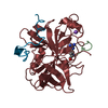
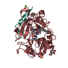
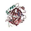
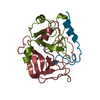
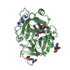
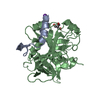
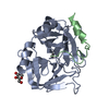
- PDB-2v9z: Structure of the Rhodococcus haloalkane dehalogenase mutant with ... -
+ Open data

- Basic information
| Entry | Database: PDB / ID: 2v9z | ||||||
|---|---|---|---|---|---|---|---|
| Title | Structure of the Rhodococcus haloalkane dehalogenase mutant with enhanced enantioselectivity | ||||||
 Components Components | HALOALKANE DEHALOGENASE | ||||||
 Keywords Keywords | HYDROLASE / PLASMID / DETOXIFICATION | ||||||
| Function / homology |  Function and homology informationhaloalkane dehalogenase / haloalkane dehalogenase activity / response to toxic substance Function and homology informationhaloalkane dehalogenase / haloalkane dehalogenase activity / response to toxic substanceSimilarity search - Function | ||||||
| Biological species |  RHODOCOCCUS RHODOCHROUS (bacteria) RHODOCOCCUS RHODOCHROUS (bacteria) | ||||||
| Method | X-RAY DIFFRACTION / MOLECULAR REPLACEMENT / Resolution: 3 Å | ||||||
 Authors Authors | Koudelakova, T. / Prokop, Z. / Sato, Y. / Lapkouski, M. / Chovancova, E. / Monincova, M. / Jesenska, A. / Emmer, J. / Senda, T. / Nagata, Y. ...Koudelakova, T. / Prokop, Z. / Sato, Y. / Lapkouski, M. / Chovancova, E. / Monincova, M. / Jesenska, A. / Emmer, J. / Senda, T. / Nagata, Y. / Kuta Smatanova, I. / Damborsky, J. | ||||||
 Citation Citation | Journal: To be Published Title: Rational Engineering of Rhodococcus Haloalkane Dehalogenase with Enhanced Enantioselectivity Authors: Koudelakova, T. / Prokop, Z. / Sato, Y. / Lapkouski, M. / Chovancova, E. / Monincova, M. / Jesenska, A. / Emmer, J. / Senda, T. / Nagata, Y. / Kuta Smatanova, I. / Damborsky, J. | ||||||
| History |
|
- Structure visualization
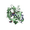
| Structure viewer | Molecule: MolmilJmol/JSmol |
|---|
- Downloads & links
-Download
| PDBx/mmCIF format | 2v9z.cif.gz | 72 KB | Display | PDBx/mmCIF format |
|---|---|---|---|---|
| PDB format | pdb2v9z.ent.gz | 53 KB | Display | PDB format |
| PDBx/mmJSON format | 2v9z.json.gz | Tree view | PDBx/mmJSON format | |
| Others |  Other downloads Other downloads |
-Validation report
| Arichive directory | https://data.pdbj.org/pub/pdb/validation_reports/v9/2v9zftp://data.pdbj.org/pub/pdb/validation_reports/v9/2v9z | HTTPS FTP |
|---|
-Related structure data
| Related structure data | 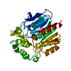 1bn6S S: Starting model for refinement |
|---|---|
| Similar structure data |


-Links
 PDBj
PDBj


- Assembly
| Deposited unit | 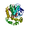
| ||||||||
|---|---|---|---|---|---|---|---|---|---|
| 1 |
| ||||||||
| Unit cell |
|
-Components
| #1: Protein | / HALOALKANE DEHALOGENASE DHAA12 Mass: 34439.059 Da / Num. of mol.: 1 / Mutation: YES Source method: isolated from a genetically manipulated source Source: (gene. exp.) RHODOCOCCUS RHODOCHROUS (bacteria) / Strain: NCIMB13064 / Production host: ESCHERICHIA COLI (E. coli) / Strain (production host): BL21(DE3) / References: UniProt: P0A3G2, haloalkane dehalogenase |
|---|---|
| Compound details | ENGINEERED RESIDUE IN CHAIN A, TRP 141 TO PHE ENGINEERED RESIDUE IN CHAIN A, PRO 142 TO ALA ...ENGINEERED |
-Experimental details
-Experiment
| Experiment | Method: X-RAY DIFFRACTION / Number of used crystals: 1 |
|---|
- Sample preparation
| Crystal | Density Matthews: 2.31 Å3/Da / Density % sol: 47 % / Description: NONE |
|---|---|
| Crystal grow | Temperature: 281 K / Method: vapor diffusion, sitting drop Details: 1 UL OF PROTEIN (5 MG/ML IN 25 MM TRIS-HCL PH 7.5, 150 MM AMMONIUM SULPHATE, 1 MM EDTA) WAS MIXED 1:1 WITH THE RESERVOIR (1 ML) CONSISTED OF 20 % PEG 6000, 0.1 M SODIUM ACETATE, 0.2 M ...Details: 1 UL OF PROTEIN (5 MG/ML IN 25 MM TRIS-HCL PH 7.5, 150 MM AMMONIUM SULPHATE, 1 MM EDTA) WAS MIXED 1:1 WITH THE RESERVOIR (1 ML) CONSISTED OF 20 % PEG 6000, 0.1 M SODIUM ACETATE, 0.2 M AMMONIUM SULPHATE, 0.1 M TRIS-HCL PH 8.5-9.0. SITTING-DROP 281K. |
-Data collection
| Diffraction | Mean temperature: 120 K |
|---|---|
| Diffraction source | Source: ROTATING ANODE / Type: ENRAF-NONIUS FR591 / Wavelength: 1.5418 |
| Detector | Detector: IMAGE PLATE / Date: Apr 21, 2007 / Details: MIRRORS |
| Radiation | Monochromator: MIRRORS / Protocol: SINGLE WAVELENGTH / Monochromatic (M) / Laue (L): M / Scattering type: x-ray |
| Radiation wavelength | Wavelength: 1.5418 Å / Relative weight: 1 |
| Reflection | Resolution: 3→53.45 Å / Num. obs: 6389 / % possible obs: 97.9 % / Observed criterion σ(I): 3.7 / Redundancy: 3.4 % / Rmerge(I) obs: 0.14 / Net I/σ(I): 8.2 |
| Reflection shell | Resolution: 3→3.08 Å / Redundancy: 3.1 % / Rmerge(I) obs: 0.43 / Mean I/σ(I) obs: 2.3 / % possible all: 92.5 |
- Processing
| Software |
| ||||||||||||||||||||||||||||||||||||||||||||||||||||||||||||||||||||||||||||||||||||||||||||||||||||||||||||||||||||||||||||||||||||||||||||||||||||||||||||||||||||||||||||||||||||||
|---|---|---|---|---|---|---|---|---|---|---|---|---|---|---|---|---|---|---|---|---|---|---|---|---|---|---|---|---|---|---|---|---|---|---|---|---|---|---|---|---|---|---|---|---|---|---|---|---|---|---|---|---|---|---|---|---|---|---|---|---|---|---|---|---|---|---|---|---|---|---|---|---|---|---|---|---|---|---|---|---|---|---|---|---|---|---|---|---|---|---|---|---|---|---|---|---|---|---|---|---|---|---|---|---|---|---|---|---|---|---|---|---|---|---|---|---|---|---|---|---|---|---|---|---|---|---|---|---|---|---|---|---|---|---|---|---|---|---|---|---|---|---|---|---|---|---|---|---|---|---|---|---|---|---|---|---|---|---|---|---|---|---|---|---|---|---|---|---|---|---|---|---|---|---|---|---|---|---|---|---|---|---|---|
| Refinement | Method to determine structure: MOLECULAR REPLACEMENT Starting model: PDB ENTRY 1BN6 Resolution: 3→53.45 Å / Cor.coef. Fo:Fc: 0.915 / Cor.coef. Fo:Fc free: 0.796 / SU B: 22.618 / SU ML: 0.422 / Cross valid method: THROUGHOUT / ESU R Free: 0.602 / Stereochemistry target values: MAXIMUM LIKELIHOOD Details: HYDROGENS HAVE BEEN ADDED IN THE RIDING POSITIONS. RESIDUES MET A 1 AND SER A 2 ARE DISORDERED. GLU A 3 DUE TO DISORDERED SIDE CHAIN IS SUBSTITUTED BY ALA. SIDE CHAINS OF ASP A 167 AND VAL A ...Details: HYDROGENS HAVE BEEN ADDED IN THE RIDING POSITIONS. RESIDUES MET A 1 AND SER A 2 ARE DISORDERED. GLU A 3 DUE TO DISORDERED SIDE CHAIN IS SUBSTITUTED BY ALA. SIDE CHAINS OF ASP A 167 AND VAL A 168 WERE MODELLED STEREOCHEMICALLY
| ||||||||||||||||||||||||||||||||||||||||||||||||||||||||||||||||||||||||||||||||||||||||||||||||||||||||||||||||||||||||||||||||||||||||||||||||||||||||||||||||||||||||||||||||||||||
| Solvent computation | Ion probe radii: 0.8 Å / Shrinkage radii: 0.8 Å / VDW probe radii: 1.2 Å / Solvent model: MASK | ||||||||||||||||||||||||||||||||||||||||||||||||||||||||||||||||||||||||||||||||||||||||||||||||||||||||||||||||||||||||||||||||||||||||||||||||||||||||||||||||||||||||||||||||||||||
| Displacement parameters | Biso mean: 15.46 Å2 | ||||||||||||||||||||||||||||||||||||||||||||||||||||||||||||||||||||||||||||||||||||||||||||||||||||||||||||||||||||||||||||||||||||||||||||||||||||||||||||||||||||||||||||||||||||||
| Refinement step | Cycle: LAST / Resolution: 3→53.45 Å
| ||||||||||||||||||||||||||||||||||||||||||||||||||||||||||||||||||||||||||||||||||||||||||||||||||||||||||||||||||||||||||||||||||||||||||||||||||||||||||||||||||||||||||||||||||||||
| Refine LS restraints |
|