 Movie
Movie Controller

[English] 日本語
 Yorodumi
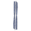
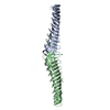
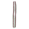
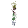
- PDB-3qc7: The structure of bacteriophage phi29 head fibers has a supercoile... -
+ Open data

- Basic information
| Entry | Database: PDB / ID: 3qc7 | ||||||
|---|---|---|---|---|---|---|---|
| Title | The structure of bacteriophage phi29 head fibers has a supercoiled triple repeating helix-turn-helix motif | ||||||
 Components Components | Head fiber protein | ||||||
 Keywords Keywords |  VIRAL PROTEIN / supercoiled triple repeating helix-turn-helix VIRAL PROTEIN / supercoiled triple repeating helix-turn-helix | ||||||
| Function / homology |  Function and homology informationviral capsid, fiber / viral procapsid / symbiont entry into host cell / virion attachment to host cell / identical protein binding Function and homology informationviral capsid, fiber / viral procapsid / symbiont entry into host cell / virion attachment to host cell / identical protein bindingSimilarity search - Function | ||||||
| Biological species |   Bacillus phage phi29 (virus) Bacillus phage phi29 (virus) | ||||||
| Method | X-RAY DIFFRACTION / SYNCHROTRON / SAD / Resolution: 1.52 Å | ||||||
 Authors Authors | Xiang, Y. / Rossmann, M.G. | ||||||
 Citation Citation | Journal: Proc.Natl.Acad.Sci.USA / Year: 2011 Title: Structure of bacteriophage {phi}29 head fibers has a supercoiled triple repeating helix-turn-helix motif. Authors: Xiang, Y. / Rossmann, M.G. | ||||||
| History |
|
- Structure visualization
| Structure viewer | Molecule: MolmilJmol/JSmol |
|---|
- Downloads & links
-Download
| PDBx/mmCIF format | 3qc7.cif.gz | 46.4 KB | Display | PDBx/mmCIF format |
|---|---|---|---|---|
| PDB format | pdb3qc7.ent.gz | 33.4 KB | Display | PDB format |
| PDBx/mmJSON format | 3qc7.json.gz | Tree view | PDBx/mmJSON format | |
| Others |  Other downloads Other downloads |
-Validation report
| Arichive directory | https://data.pdbj.org/pub/pdb/validation_reports/qc/3qc7ftp://data.pdbj.org/pub/pdb/validation_reports/qc/3qc7 | HTTPS FTP |
|---|
-Related structure data
| Similar structure data |
|---|

-Links
 PDBj
PDBj- Assembly
| Deposited unit | 
| ||||||||
|---|---|---|---|---|---|---|---|---|---|
| 1 | 
| ||||||||
| Unit cell |
|
-Components
| #1: Protein | Mass: 18923.359 Da / Num. of mol.: 1 / Fragment: gp8.5 fibrous portion (UNP residues 110-280) Source method: isolated from a genetically manipulated source Source: (gene. exp.) Bacillus phage phi29 (virus) / Gene: 8.5 / Plasmid: pET30b / Production host:  Escherichia coli (E. coli) / References: UniProt: P20344, UniProt: B3VMP4*PLUS Escherichia coli (E. coli) / References: UniProt: P20344, UniProt: B3VMP4*PLUS |
|---|---|
| #2: Water | ChemComp-HOH / Water Mass: 18.015 Da / Num. of mol.: 179 / Source method: isolated from a natural source / Formula: H2O Mass: 18.015 Da / Num. of mol.: 179 / Source method: isolated from a natural source / Formula: H2O |
-Experimental details
-Experiment
| Experiment | Method: X-RAY DIFFRACTION / Number of used crystals: 1 |
|---|
- Sample preparation
| Crystal | Density Matthews: 2.4 Å3/Da / Density % sol: 48.67 % |
|---|---|
| Crystal grow | Temperature: 298 K / Method: vapor diffusion, hanging drop / pH: 6.8 Details: The gp8.5 fragment crystals were grown in 100mM sodium phosphate at pH ~6.8 and 8% w/v PEG 3000. It took about one week for crystals to appear, VAPOR DIFFUSION, HANGING DROP, temperature 298K |
-Data collection
| Diffraction | Mean temperature: 100 K |
|---|---|
| Diffraction source | Source: SYNCHROTRON / Site: APS  / Beamline: 23-ID-D / Wavelength: 0.968 Å / Beamline: 23-ID-D / Wavelength: 0.968 Å |
| Detector | Type: MARMOSAIC 300 mm CCD / Detector: CCD / Date: Jan 1, 2010 |
| Radiation | Monochromator: Si 111 / Protocol: SINGLE WAVELENGTH / Monochromatic (M) / Laue (L): M / Scattering type: x-ray |
| Radiation wavelength | Wavelength: 0.968 Å / Relative weight: 1 |
| Reflection | Resolution: 1.52→70 Å / Num. all: 28649 / Num. obs: 28649 / % possible obs: 98.6 % / Observed criterion σ(F): -3 / Observed criterion σ(I): -3 / Rmerge(I) obs: 0.07 / Rsym value: 0.07 / Net I/σ(I): 33.4 |
| Reflection shell | Resolution: 1.52→1.57 Å / Rmerge(I) obs: 0.407 / Mean I/σ(I) obs: 4.6 / % possible all: 97.7 |
- Processing
| Software |
| |||||||||||||||||||||||||||||||||||||||||||||||||||||||||||||||||
|---|---|---|---|---|---|---|---|---|---|---|---|---|---|---|---|---|---|---|---|---|---|---|---|---|---|---|---|---|---|---|---|---|---|---|---|---|---|---|---|---|---|---|---|---|---|---|---|---|---|---|---|---|---|---|---|---|---|---|---|---|---|---|---|---|---|---|
| Refinement | Method to determine structure: SAD / Resolution: 1.52→29.47 Å / Cor.coef. Fo:Fc: 0.956 / Cor.coef. Fo:Fc free: 0.946 / SU B: 1.121 / SU ML: 0.043 / Cross valid method: THROUGHOUT / σ(F): 0 / σ(I): 0 / ESU R Free: 0.077 Stereochemistry target values: MAXIMUM LIKELIHOOD WITH PHASES Details: HYDROGENS HAVE BEEN ADDED IN THE RIDING POSITIONS
| |||||||||||||||||||||||||||||||||||||||||||||||||||||||||||||||||
| Solvent computation | Ion probe radii: 0.8 Å / Shrinkage radii: 0.8 Å / VDW probe radii: 1.4 Å / Solvent model: MASK | |||||||||||||||||||||||||||||||||||||||||||||||||||||||||||||||||
| Displacement parameters | Biso mean: 22.08 Å2
| |||||||||||||||||||||||||||||||||||||||||||||||||||||||||||||||||
| Refinement step | Cycle: LAST / Resolution: 1.52→29.47 Å
| |||||||||||||||||||||||||||||||||||||||||||||||||||||||||||||||||
| Refine LS restraints |
| |||||||||||||||||||||||||||||||||||||||||||||||||||||||||||||||||
| LS refinement shell | Resolution: 1.521→1.56 Å / Total num. of bins used: 20
|
