 Movie
Movie Controller

[English] 日本語
 Yorodumi
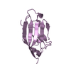
- PDB-3q2c: Binding properties to HLA class I molecules and the structure of ... -
+ Open data

- Basic information
| Entry | Database: PDB / ID: 3q2c | ||||||
|---|---|---|---|---|---|---|---|
| Title | Binding properties to HLA class I molecules and the structure of the leukocyte Ig-like receptor A3 (LILRA3/ILT6/LIR4/CD85e) | ||||||
 Components Components | Leukocyte immunoglobulin-like receptor subfamily A member 3 | ||||||
 Keywords Keywords |  IMMUNE SYSTEM / LILRA3 / ILT6 / Activating receptor / HLA binding IMMUNE SYSTEM / LILRA3 / ILT6 / Activating receptor / HLA binding | ||||||
| Function / homology |  Function and homology information Function and homology informationneutrophil degranulation / tertiary granule membrane / ficolin-1-rich granule membrane / specific granule membrane / antigen binding / defense response / Immunoregulatory interactions between a Lymphoid and a non-Lymphoid cell / signaling receptor activity / adaptive immune response / Neutrophil degranulation ...neutrophil degranulation / tertiary granule membrane / ficolin-1-rich granule membrane / specific granule membrane / antigen binding / defense response / Immunoregulatory interactions between a Lymphoid and a non-Lymphoid cell / signaling receptor activity / adaptive immune response / Neutrophil degranulation / signal transduction / extracellular region / plasma membraneSimilarity search - Function | ||||||
| Biological species |  Homo sapiens (human) Homo sapiens (human) | ||||||
| Method | X-RAY DIFFRACTION / MOLECULAR REPLACEMENT / Resolution: 2.5 Å | ||||||
 Authors Authors | Ryu, M. / Chen, Y. / Qi, J.X. / Liu, J. / Shi, Y. / Cheng, H. / Gao, G.F. | ||||||
 Citation Citation | Journal: Plos One / Year: 2011 Title: LILRA3 binds both classical and non-classical HLA class I molecules but with reduced affinities compared to LILRB1/LILRB2: structural evidence Authors: Ryu, M. / Chen, Y. / Qi, J. / Liu, J. / Fan, Z. / Nam, G. / Shi, Y. / Cheng, H. / Gao, G.F. | ||||||
| History |
|
- Structure visualization
| Structure viewer | Molecule: MolmilJmol/JSmol |
|---|
- Downloads & links
-Download
| PDBx/mmCIF format | 3q2c.cif.gz | 51.6 KB | Display | PDBx/mmCIF format |
|---|---|---|---|---|
| PDB format | pdb3q2c.ent.gz | 36.8 KB | Display | PDB format |
| PDBx/mmJSON format | 3q2c.json.gz | Tree view | PDBx/mmJSON format | |
| Others |  Other downloads Other downloads |
-Validation report
| Arichive directory | https://data.pdbj.org/pub/pdb/validation_reports/q2/3q2cftp://data.pdbj.org/pub/pdb/validation_reports/q2/3q2c | HTTPS FTP |
|---|
-Related structure data
| Related structure data |  1vdgS S: Starting model for refinement |
|---|---|
| Similar structure data |


-Links
 PDBj
PDBj
- Assembly
| Deposited unit | 
| ||||||||
|---|---|---|---|---|---|---|---|---|---|
| 1 |
| ||||||||
| Unit cell |
|
-Components
| #1: Protein | Mass: 10792.341 Da / Num. of mol.: 1 / Fragment: N-TERMINAL DOMAIN, UNP residues 24-120 / Source method: isolated from a natural source / Source: (natural) Homo sapiens (human) / References: UniProt: Q8N6C8 |
|---|---|
| #2: Water | ChemComp-HOH / Water Mass: 18.015 Da / Num. of mol.: 32 / Source method: isolated from a natural source / Formula: H2O Mass: 18.015 Da / Num. of mol.: 32 / Source method: isolated from a natural source / Formula: H2O |
| Sequence details | THE SEQUENCE IS BASED ON REFERENCE 3 OF DATABASE Q8N6C8 (LIRA3_HUMAN). RESIDUE VAL 82 (UNP RESIDUE ...THE SEQUENCE IS BASED ON REFERENCE 3 OF DATABASE Q8N6C8 (LIRA3_HUMAN). RESIDUE VAL 82 (UNP RESIDUE NUMBER 105) IS A SEQUENCE CONFLICT. |
-Experimental details
-Experiment
| Experiment | Method: X-RAY DIFFRACTION / Number of used crystals: 1 |
|---|
- Sample preparation
| Crystal | Density Matthews: 2.41 Å3/Da / Density % sol: 48.91 % |
|---|---|
| Crystal grow | Temperature: 291 K / Method: vapor diffusion, hanging drop / pH: 7.5 Details: 1.0M ammonium sulfate, 0.1M HEPES buffer (pH 7.5), 0.75% (w/v) PEG 8000, VAPOR DIFFUSION, HANGING DROP, temperature 291K |
-Data collection
| Diffraction | Mean temperature: 100 K |
|---|---|
| Diffraction source | Source: ROTATING ANODE / Type: RIGAKU MICROMAX-007 HF / Wavelength: 1.5418 Å |
| Detector | Type: RIGAKU RAXIS IV++ / Detector: IMAGE PLATE / Date: Dec 20, 2009 / Details: mirrors |
| Radiation | Monochromator: GRAPHITE / Protocol: SINGLE WAVELENGTH / Monochromatic (M) / Laue (L): M / Scattering type: x-ray |
| Radiation wavelength | Wavelength: 1.5418 Å / Relative weight: 1 |
| Reflection | Resolution: 2.5→50 Å / Num. obs: 3558 / % possible obs: 96.7 % / Observed criterion σ(F): 2 / Observed criterion σ(I): 2 |
| Reflection shell | Resolution: 2.5→2.59 Å / % possible all: 92.5 |
- Processing
| Software |
| ||||||||||||||||||||||||||||||||||||||||
|---|---|---|---|---|---|---|---|---|---|---|---|---|---|---|---|---|---|---|---|---|---|---|---|---|---|---|---|---|---|---|---|---|---|---|---|---|---|---|---|---|---|
| Refinement | Method to determine structure: MOLECULAR REPLACEMENT Starting model: PDB ENTRY 1VDG Resolution: 2.5→23.947 Å / SU ML: 0.18 / σ(F): 0.16 / Phase error: 29.01 / Stereochemistry target values: ML
| ||||||||||||||||||||||||||||||||||||||||
| Solvent computation | Shrinkage radii: 0.9 Å / VDW probe radii: 1.11 Å / Solvent model: FLAT BULK SOLVENT MODEL / Bsol: 42.554 Å2 / ksol: 0.362 e/Å3 | ||||||||||||||||||||||||||||||||||||||||
| Displacement parameters |
| ||||||||||||||||||||||||||||||||||||||||
| Refinement step | Cycle: LAST / Resolution: 2.5→23.947 Å
| ||||||||||||||||||||||||||||||||||||||||
| Refine LS restraints |
| ||||||||||||||||||||||||||||||||||||||||
| LS refinement shell |
| ||||||||||||||||||||||||||||||||||||||||
| Refinement TLS params. | Method: refined / Origin x: 22.2116 Å / Origin y: 1.8417 Å / Origin z: 14.1459 Å
| ||||||||||||||||||||||||||||||||||||||||
| Refinement TLS group | Selection details: all |
