 Movie
Movie Controller


+ Open data

- Basic information
| Entry | Database: PDB / ID: 2i5s | |||||||||
|---|---|---|---|---|---|---|---|---|---|---|
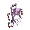
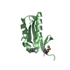
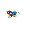
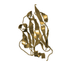
| Title | Crystal structure of onconase with bound nucleic acid | |||||||||
 Components Components |
| |||||||||
 Keywords Keywords | HYDROLASE/DNA /  Onconase / P-30 protein / ribonuclease / anti-tumor / Structural Genomics / PSI-2 / Protein Structure Initiative / Center for Eukaryotic Structural Genomics / CESG / HYDROLASE-DNA COMPLEX Onconase / P-30 protein / ribonuclease / anti-tumor / Structural Genomics / PSI-2 / Protein Structure Initiative / Center for Eukaryotic Structural Genomics / CESG / HYDROLASE-DNA COMPLEX | |||||||||
| Function / homology |  Function and homology informationHydrolases; Acting on ester bonds; Endoribonucleases producing 3'-phosphomonoesters / endonuclease activity / nucleic acid binding Function and homology informationHydrolases; Acting on ester bonds; Endoribonucleases producing 3'-phosphomonoesters / endonuclease activity / nucleic acid bindingSimilarity search - Function | |||||||||
| Biological species | Rana pipiens (northern leopard frog) | |||||||||
| Method | X-RAY DIFFRACTION / Resolution: 1.9 Å | |||||||||
 Authors Authors | Bae, E. / Lee, J.E. / Raines, R.T. / Wesenberg, G.E. / Phillips Jr., G.N. / Bitto, E. / Bingman, C.A. / Center for Eukaryotic Structural Genomics (CESG) | |||||||||
 Citation Citation | Journal: J.Mol.Biol. / Year: 2008 Title: Structural basis for catalysis by onconase. Authors: Lee, J.E. / Bae, E. / Bingman, C.A. / Phillips Jr., G.N. / Raines, R.T. | |||||||||
| History |
|
- Structure visualization
| Structure viewer | Molecule: MolmilJmol/JSmol |
|---|
- Downloads & links
-Download
| PDBx/mmCIF format | 2i5s.cif.gz | 42.2 KB | Display | PDBx/mmCIF format |
|---|---|---|---|---|
| PDB format | pdb2i5s.ent.gz | 26 KB | Display | PDB format |
| PDBx/mmJSON format | 2i5s.json.gz | Tree view | PDBx/mmJSON format | |
| Others |  Other downloads Other downloads |
-Validation report
| Arichive directory | https://data.pdbj.org/pub/pdb/validation_reports/i5/2i5sftp://data.pdbj.org/pub/pdb/validation_reports/i5/2i5s | HTTPS FTP |
|---|
-Related structure data
| Related structure data |  2gmkC  1oncS C: citing same article ( S: Starting model for refinement |
|---|---|
| Similar structure data | |
| Other databases |


-Links
 PDBj
PDBj

- Assembly
| Deposited unit | 
| ||||||||
|---|---|---|---|---|---|---|---|---|---|
| 1 |
| ||||||||
| Unit cell |
| ||||||||
| Components on special symmetry positions |
|
-Components
| #1: Protein | Mass: 11845.648 Da / Num. of mol.: 1 Source method: isolated from a genetically manipulated source Source: (gene. exp.) Rana pipiens (northern leopard frog) / Gene: RNP30_RANPI / Plasmid: pET 22b(+) / Species (production host): Escherichia coli / Production host:  Escherichia coli BL21(DE3) (bacteria) / Strain (production host): BL21(DE3) Escherichia coli BL21(DE3) (bacteria) / Strain (production host): BL21(DE3)References: UniProt: P22069, Hydrolases; Acting on ester bonds; Endoribonucleases producing 3'-phosphomonoesters |
|---|---|
| #2: DNA chain | Mass: 1200.829 Da / Num. of mol.: 1 / Source method: obtained synthetically |
| #3: Water | ChemComp-HOH / Water Mass: 18.015 Da / Num. of mol.: 155 / Source method: isolated from a natural source / Formula: H2O Mass: 18.015 Da / Num. of mol.: 155 / Source method: isolated from a natural source / Formula: H2O |
-Experimental details
-Experiment
| Experiment | Method: X-RAY DIFFRACTION / Number of used crystals: 1 |
|---|
- Sample preparation
| Crystal | Density Matthews: 1.9 Å3/Da / Density % sol: 41.186596 % | ||||||||||||||||||||||||||||||||||||
|---|---|---|---|---|---|---|---|---|---|---|---|---|---|---|---|---|---|---|---|---|---|---|---|---|---|---|---|---|---|---|---|---|---|---|---|---|---|
| Crystal grow | Temperature: 293 K / Method: vapor diffusion, hanging drop Details: PROTEIN SOLUTION (10 MG/ML PROTEIN, 0.003 M SINGLE-STRANDED DNA OLIGOMER) MIXED IN A 1:1 RATIO WITH WELL SOLUTION (25% PEG 3350, 0.1 M HEPES PH 7.5) CRYO-PROTECTED WITH WELL SOLUTION ...Details: PROTEIN SOLUTION (10 MG/ML PROTEIN, 0.003 M SINGLE-STRANDED DNA OLIGOMER) MIXED IN A 1:1 RATIO WITH WELL SOLUTION (25% PEG 3350, 0.1 M HEPES PH 7.5) CRYO-PROTECTED WITH WELL SOLUTION SUPPLEMENTED WITH 20% ETHYLENE GLYCOL, VAPOR DIFFUSION, HANGING DROP, temperature 293K | ||||||||||||||||||||||||||||||||||||
| Components of the solutions |
|
-Data collection
| Diffraction | Mean temperature: 100 K | ||||||||||||||||||||||||||||||||||||||||||||||||||||||||||||||||||||||||||||||||||||||||||||||||||||||
|---|---|---|---|---|---|---|---|---|---|---|---|---|---|---|---|---|---|---|---|---|---|---|---|---|---|---|---|---|---|---|---|---|---|---|---|---|---|---|---|---|---|---|---|---|---|---|---|---|---|---|---|---|---|---|---|---|---|---|---|---|---|---|---|---|---|---|---|---|---|---|---|---|---|---|---|---|---|---|---|---|---|---|---|---|---|---|---|---|---|---|---|---|---|---|---|---|---|---|---|---|---|---|---|
| Diffraction source | Source: ROTATING ANODE / Type: BRUKER AXS MICROSTAR / Wavelength: 1.5418 Å | ||||||||||||||||||||||||||||||||||||||||||||||||||||||||||||||||||||||||||||||||||||||||||||||||||||||
| Detector | Type: BRUKER SMART 6000 / Detector: CCD / Date: May 2, 2006 / Details: MONTEL OPTICS | ||||||||||||||||||||||||||||||||||||||||||||||||||||||||||||||||||||||||||||||||||||||||||||||||||||||
| Radiation | Monochromator: Graded Multilayer / Protocol: SINGLE WAVELENGTH / Scattering type: x-ray | ||||||||||||||||||||||||||||||||||||||||||||||||||||||||||||||||||||||||||||||||||||||||||||||||||||||
| Radiation wavelength | Wavelength: 1.5418 Å / Relative weight: 1 | ||||||||||||||||||||||||||||||||||||||||||||||||||||||||||||||||||||||||||||||||||||||||||||||||||||||
| Reflection | Resolution: 1.9→64.6145 Å / Num. obs: 9270 / % possible obs: 99.7 % / Redundancy: 9.84 % / Rmerge(I) obs: 0.1249 / Net I/σ(I): 12.3 | ||||||||||||||||||||||||||||||||||||||||||||||||||||||||||||||||||||||||||||||||||||||||||||||||||||||
| Reflection shell |
|
-Phasing
| Phasing MR | Rfactor: 0.569 / Cor.coef. Fo:Fc: 0.23
|
|---|
- Processing
| Software |
| |||||||||||||||||||||||||||||||||||||||||||||||||||||||||||||||||||||||||||||||||||||||||||||||||||||||||||||||||||||||||||||||||||||||||||||||||||
|---|---|---|---|---|---|---|---|---|---|---|---|---|---|---|---|---|---|---|---|---|---|---|---|---|---|---|---|---|---|---|---|---|---|---|---|---|---|---|---|---|---|---|---|---|---|---|---|---|---|---|---|---|---|---|---|---|---|---|---|---|---|---|---|---|---|---|---|---|---|---|---|---|---|---|---|---|---|---|---|---|---|---|---|---|---|---|---|---|---|---|---|---|---|---|---|---|---|---|---|---|---|---|---|---|---|---|---|---|---|---|---|---|---|---|---|---|---|---|---|---|---|---|---|---|---|---|---|---|---|---|---|---|---|---|---|---|---|---|---|---|---|---|---|---|---|---|---|---|
| Refinement | Starting model: PDB ENTRY 1ONC Resolution: 1.9→32.49 Å / Cor.coef. Fo:Fc: 0.947 / Cor.coef. Fo:Fc free: 0.912 / WRfactor Rfree: 0.211 / WRfactor Rwork: 0.165 / SU B: 3.248 / SU ML: 0.098 / Cross valid method: THROUGHOUT / σ(F): 0 / ESU R: 0.161 / ESU R Free: 0.158 / Stereochemistry target values: MAXIMUM LIKELIHOOD / Details: HYDROGENS HAVE BEEN ADDED IN THE RIDING POSITIONS
| |||||||||||||||||||||||||||||||||||||||||||||||||||||||||||||||||||||||||||||||||||||||||||||||||||||||||||||||||||||||||||||||||||||||||||||||||||
| Solvent computation | Ion probe radii: 0.8 Å / Shrinkage radii: 0.8 Å / VDW probe radii: 1.2 Å / Solvent model: BABINET MODEL PLUS MASK | |||||||||||||||||||||||||||||||||||||||||||||||||||||||||||||||||||||||||||||||||||||||||||||||||||||||||||||||||||||||||||||||||||||||||||||||||||
| Displacement parameters | Biso mean: 16.602 Å2
| |||||||||||||||||||||||||||||||||||||||||||||||||||||||||||||||||||||||||||||||||||||||||||||||||||||||||||||||||||||||||||||||||||||||||||||||||||
| Refinement step | Cycle: LAST / Resolution: 1.9→32.49 Å
| |||||||||||||||||||||||||||||||||||||||||||||||||||||||||||||||||||||||||||||||||||||||||||||||||||||||||||||||||||||||||||||||||||||||||||||||||||
| Refine LS restraints |
| |||||||||||||||||||||||||||||||||||||||||||||||||||||||||||||||||||||||||||||||||||||||||||||||||||||||||||||||||||||||||||||||||||||||||||||||||||
| LS refinement shell | Refine-ID: X-RAY DIFFRACTION / Total num. of bins used: 20
|
