 Movie
Movie Controller

[English] 日本語
 Yorodumi
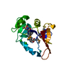
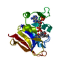
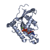
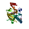
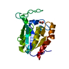
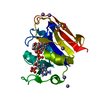
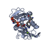
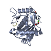
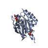
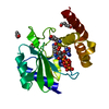
- PDB-3q1h: Crystal Structure of Dihydrofolate Reductase from Yersinia pestis -
+ Open data

- Basic information
| Entry | Database: PDB / ID: 3q1h | ||||||
|---|---|---|---|---|---|---|---|
| Title | Crystal Structure of Dihydrofolate Reductase from Yersinia pestis | ||||||
 Components Components | Dihydrofolate reductase | ||||||
 Keywords Keywords | OXIDOREDUCTASE / Structural Genomics / Center for Structural Genomics of Infectious Diseases / CSGID / alpha beta fold / cytosol | ||||||
| Function / homology |  Function and homology information Function and homology informationdihydrofolate metabolic process / glycine biosynthetic process / dihydrofolate reductase / folic acid metabolic process / dihydrofolate reductase activity / tetrahydrofolate biosynthetic process / one-carbon metabolic process / NADP binding / metal ion binding / cytosolSimilarity search - Function | ||||||
| Biological species |   Yersinia pestis (bacteria) Yersinia pestis (bacteria) | ||||||
| Method | X-RAY DIFFRACTION / SYNCHROTRON / MOLECULAR REPLACEMENT / Resolution: 1.804 Å | ||||||
 Authors Authors | Maltseva, N. / Kim, Y. / Makowska-Grzyska, M. / Mulligan, R. / Papazisi, L. / Anderson, W.F. / Joachimiak, A. / Center for Structural Genomics of Infectious Diseases (CSGID) | ||||||
 Citation Citation | Journal: To be Published Title: Crystal Structure of Dihydrofolate Reductase from Yersinia pestis Authors: Maltseva, N. / Kim, Y. / Makowska-Grzyska, M. / Mulligan, R. / Papazisi, L. / Anderson, W.F. / Joachimiak, A. / Center for Structural Genomics of Infectious Diseases (CSGID) | ||||||
| History |
|
- Structure visualization
| Structure viewer | Molecule: MolmilJmol/JSmol |
|---|
- Downloads & links
-Download
| PDBx/mmCIF format | 3q1h.cif.gz | 82.2 KB | Display | PDBx/mmCIF format |
|---|---|---|---|---|
| PDB format | pdb3q1h.ent.gz | 60.9 KB | Display | PDB format |
| PDBx/mmJSON format | 3q1h.json.gz | Tree view | PDBx/mmJSON format | |
| Others |  Other downloads Other downloads |
-Validation report
| Arichive directory | https://data.pdbj.org/pub/pdb/validation_reports/q1/3q1hftp://data.pdbj.org/pub/pdb/validation_reports/q1/3q1h | HTTPS FTP |
|---|
-Related structure data
| Related structure data | 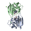 1ddsS S: Starting model for refinement |
|---|---|
| Similar structure data | |
| Other databases |
-Links
 PDBj
PDBj
- Assembly
| Deposited unit | 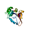
| ||||||||||||
|---|---|---|---|---|---|---|---|---|---|---|---|---|---|
| 1 |
| ||||||||||||
| Unit cell |
| ||||||||||||
| Components on special symmetry positions |
|
-Components
| #1: Protein | / Dihydrofolate reductase type I Mass: 18350.652 Da / Num. of mol.: 1 Source method: isolated from a genetically manipulated source Source: (gene. exp.) Yersinia pestis (bacteria) / Strain: CO92 / Gene: folA, y3688, YPO0486, YP_3693 / Plasmid: pMCSG7 / Production host: Escherichia coli (E. coli) / Strain (production host): KRXpGro7References: UniProt: Q7CG83, UniProt: A0A3N4BLI0*PLUS, dihydrofolate reductase | ||
|---|---|---|---|
| #2: Chemical | Sulfate  Mass: 96.063 Da / Num. of mol.: 2 / Source method: obtained synthetically / Formula: SO4 Mass: 96.063 Da / Num. of mol.: 2 / Source method: obtained synthetically / Formula: SO4#3: Water | ChemComp-HOH / | Water Mass: 18.015 Da / Num. of mol.: 163 / Source method: isolated from a natural source / Formula: H2O Mass: 18.015 Da / Num. of mol.: 163 / Source method: isolated from a natural source / Formula: H2O |
-Experimental details
-Experiment
| Experiment | Method: X-RAY DIFFRACTION / Number of used crystals: 1 |
|---|
- Sample preparation
| Crystal | Density Matthews: 2.11 Å3/Da / Density % sol: 41.68 % |
|---|---|
| Crystal grow | Temperature: 289 K / Method: vapor diffusion, sitting drop / pH: 5.5 Details: 0.2M Lithium Sulfate, 0.1M Bis-Tris pH5.5, 25% PEG 3350, VAPOR DIFFUSION, SITTING DROP, temperature 289K |
-Data collection
| Diffraction | Mean temperature: 100 K |
|---|---|
| Diffraction source | Source: SYNCHROTRON / Site: APS  / Beamline: 19-ID / Wavelength: 0.9793 Å / Beamline: 19-ID / Wavelength: 0.9793 Å |
| Detector | Type: ADSC QUANTUM 315r / Detector: CCD / Date: Dec 15, 2010 / Details: mirrors |
| Radiation | Monochromator: double crystal monochromator / Protocol: SINGLE WAVELENGTH / Monochromatic (M) / Laue (L): M / Scattering type: x-ray |
| Radiation wavelength | Wavelength: 0.9793 Å / Relative weight: 1 |
| Reflection | Resolution: 1.8→50 Å / Num. all: 14518 / Num. obs: 14518 / % possible obs: 99 % / Observed criterion σ(F): 0 / Observed criterion σ(I): 0 / Redundancy: 7.6 % / Biso Wilson estimate: 18.1 Å2 / Rsym value: 0.07 / Net I/σ(I): 12.6 |
| Reflection shell | Resolution: 1.8→1.83 Å / Redundancy: 4 % / Mean I/σ(I) obs: 2.88 / Num. unique all: 640 / Rsym value: 0.393 / % possible all: 89 |
- Processing
| Software |
| ||||||||||||||||||||||||||||||||||||||||||
|---|---|---|---|---|---|---|---|---|---|---|---|---|---|---|---|---|---|---|---|---|---|---|---|---|---|---|---|---|---|---|---|---|---|---|---|---|---|---|---|---|---|---|---|
| Refinement | Method to determine structure: MOLECULAR REPLACEMENT Starting model: PDBID 1DDS Resolution: 1.804→33.759 Å / SU ML: 0.18 / Isotropic thermal model: mixed / Cross valid method: THROUGHOUT / σ(F): 0 / Stereochemistry target values: ML
| ||||||||||||||||||||||||||||||||||||||||||
| Solvent computation | Shrinkage radii: 0.83 Å / VDW probe radii: 1.1 Å / Solvent model: FLAT BULK SOLVENT MODEL / Bsol: 30.421 Å2 / ksol: 0.353 e/Å3 | ||||||||||||||||||||||||||||||||||||||||||
| Displacement parameters | Biso mean: 21.7 Å2
| ||||||||||||||||||||||||||||||||||||||||||
| Refinement step | Cycle: LAST / Resolution: 1.804→33.759 Å
| ||||||||||||||||||||||||||||||||||||||||||
| Refine LS restraints |
| ||||||||||||||||||||||||||||||||||||||||||
| LS refinement shell | Refine-ID: X-RAY DIFFRACTION
| ||||||||||||||||||||||||||||||||||||||||||
| Refinement TLS params. | Method: refined / Origin x: 15.8425 Å / Origin y: -12.2798 Å / Origin z: 13.0938 Å
| ||||||||||||||||||||||||||||||||||||||||||
| Refinement TLS group | Selection details: chain A |
