 Movie
Movie Controller

[English] 日本語
 Yorodumi
- PDB-3pyw: The structure of the SLH domain from B. anthracis surface array p... -
+ Open data

- Basic information
| Entry | Database: PDB / ID: 3pyw | ||||||
|---|---|---|---|---|---|---|---|
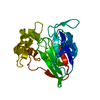
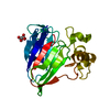
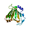
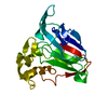
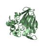
| Title | The structure of the SLH domain from B. anthracis surface array protein at 1.8A | ||||||
 Components Components | S-layer protein sap | ||||||
 Keywords Keywords | STRUCTURAL PROTEIN / SLH-domains / polysaccharide binding / GST-SLH / cell wall / Structural Genomics / PSI-Biology / Protein Structure Initiative / Midwest Center for Structural Genomics / MCSG / polysaccharide / S-layer | ||||||
| Function / homology |  Function and homology information Function and homology information | ||||||
| Biological species |  Bacillus anthracis (anthrax bacterium) Bacillus anthracis (anthrax bacterium) | ||||||
| Method | X-RAY DIFFRACTION / SYNCHROTRON / SAD / Resolution: 1.8 Å | ||||||
 Authors Authors | Zhang, R. / Wilton, R. / Kern, J. / Joachimiak, A. / Schneewind, O. / Midwest Center for Structural Genomics (MCSG) | ||||||
 Citation Citation | Journal: J.Biol.Chem. / Year: 2011 Title: Structure of Surface Layer Homology (SLH) Domains from Bacillus anthracis Surface Array Protein. Authors: Kern, J. / Wilton, R. / Zhang, R. / Binkowski, T.A. / Joachimiak, A. / Schneewind, O. | ||||||
| History |
|
- Structure visualization
| Structure viewer | Molecule: MolmilJmol/JSmol |
|---|
- Downloads & links
-Download
| PDBx/mmCIF format | 3pyw.cif.gz | 85.4 KB | Display | PDBx/mmCIF format |
|---|---|---|---|---|
| PDB format | pdb3pyw.ent.gz | 64.6 KB | Display | PDB format |
| PDBx/mmJSON format | 3pyw.json.gz | Tree view | PDBx/mmJSON format | |
| Others |  Other downloads Other downloads |
-Validation report
| Arichive directory | https://data.pdbj.org/pub/pdb/validation_reports/py/3pywftp://data.pdbj.org/pub/pdb/validation_reports/py/3pyw | HTTPS FTP |
|---|
-Related structure data
| Similar structure data | |
|---|---|
| Other databases |


-Links
 PDBj
PDBj- Assembly
| Deposited unit | 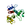
| |||||||||
|---|---|---|---|---|---|---|---|---|---|---|
| 1 |
| |||||||||
| Unit cell |
| |||||||||
| Components on special symmetry positions |
|
-Components
| #1: Protein | / Surface array protein / Surface layer protein Mass: 21916.781 Da / Num. of mol.: 1 / Fragment: SLH 1, SLH 2, and SLH 3 domains Source method: isolated from a genetically manipulated source Source: (gene. exp.) Bacillus anthracis (anthrax bacterium) / Strain: steme 34F2 / Gene: BAS0841, BA_0885, GBAA_0885, GI:49183865, sap / Plasmid: pET16b / Production host: Escherichia coli (E. coli) / Strain (production host): BL21(DE3) / References: UniProt: P49051 |
|---|---|
| #2: Chemical | ChemComp-SO4 / Sulfate  Mass: 96.063 Da / Num. of mol.: 1 / Source method: obtained synthetically / Formula: SO4 Mass: 96.063 Da / Num. of mol.: 1 / Source method: obtained synthetically / Formula: SO4 |
| #3: Water | ChemComp-HOH / Water Mass: 18.015 Da / Num. of mol.: 241 / Source method: isolated from a natural source / Formula: H2O Mass: 18.015 Da / Num. of mol.: 241 / Source method: isolated from a natural source / Formula: H2O |
-Experimental details
-Experiment
| Experiment | Method: X-RAY DIFFRACTION / Number of used crystals: 1 |
|---|
- Sample preparation
| Crystal | Density Matthews: 3.46 Å3/Da / Density % sol: 64.48 % |
|---|---|
| Crystal grow | Temperature: 289 K / Method: vapor diffusion, sitting drop / pH: 5.5 Details: 0.1M citrate pH5.5, 2.0M ammonium sulfate, VAPOR DIFFUSION, SITTING DROP, temperature 289K |
-Data collection
| Diffraction | Mean temperature: 100 K |
|---|---|
| Diffraction source | Source: SYNCHROTRON / Site: APS  / Beamline: 19-ID / Wavelength: 0.9798 Å / Beamline: 19-ID / Wavelength: 0.9798 Å |
| Detector | Type: ADSC QUANTUM 315r / Detector: CCD / Date: Jun 27, 2008 / Details: mirrors |
| Radiation | Monochromator: Si 111 channel / Protocol: SINGLE WAVELENGTH / Monochromatic (M) / Laue (L): M / Scattering type: x-ray |
| Radiation wavelength | Wavelength: 0.9798 Å / Relative weight: 1 |
| Reflection | Resolution: 1.8→54.96 Å / Num. all: 27789 / Num. obs: 27750 / % possible obs: 99.86 % / Observed criterion σ(F): 2 / Observed criterion σ(I): 2 / Redundancy: 11.1 % / Rmerge(I) obs: 0.099 / Net I/σ(I): 26.83 |
| Reflection shell | Resolution: 1.8→1.846 Å / Redundancy: 6.3 % / Rmerge(I) obs: 0.748 / Mean I/σ(I) obs: 1.61 / Num. unique all: 2133 / % possible all: 98.55 |
- Processing
| Software |
| |||||||||||||||||||||||||||||||||||||||||||||||||||||||||||||||||||||||||||||||||||||||||||||||||||||||||||||||||||||||||||||
|---|---|---|---|---|---|---|---|---|---|---|---|---|---|---|---|---|---|---|---|---|---|---|---|---|---|---|---|---|---|---|---|---|---|---|---|---|---|---|---|---|---|---|---|---|---|---|---|---|---|---|---|---|---|---|---|---|---|---|---|---|---|---|---|---|---|---|---|---|---|---|---|---|---|---|---|---|---|---|---|---|---|---|---|---|---|---|---|---|---|---|---|---|---|---|---|---|---|---|---|---|---|---|---|---|---|---|---|---|---|---|---|---|---|---|---|---|---|---|---|---|---|---|---|---|---|---|
| Refinement | Method to determine structure: SAD / Resolution: 1.8→54.96 Å / Cor.coef. Fo:Fc: 0.961 / Cor.coef. Fo:Fc free: 0.948 / SU B: 3.211 / SU ML: 0.053 / Cross valid method: THROUGHOUT / σ(I): 1 / ESU R: 0.088 / ESU R Free: 0.087 Stereochemistry target values: MAXIMUM LIKELIHOOD WITH PHASES Details: HYDROGENS HAVE BEEN ADDED IN THE RIDING POSITIONS
| |||||||||||||||||||||||||||||||||||||||||||||||||||||||||||||||||||||||||||||||||||||||||||||||||||||||||||||||||||||||||||||
| Solvent computation | Ion probe radii: 0.8 Å / Shrinkage radii: 0.8 Å / VDW probe radii: 1.2 Å / Solvent model: MASK | |||||||||||||||||||||||||||||||||||||||||||||||||||||||||||||||||||||||||||||||||||||||||||||||||||||||||||||||||||||||||||||
| Displacement parameters | Biso mean: 20.23 Å2
| |||||||||||||||||||||||||||||||||||||||||||||||||||||||||||||||||||||||||||||||||||||||||||||||||||||||||||||||||||||||||||||
| Refinement step | Cycle: LAST / Resolution: 1.8→54.96 Å
| |||||||||||||||||||||||||||||||||||||||||||||||||||||||||||||||||||||||||||||||||||||||||||||||||||||||||||||||||||||||||||||
| Refine LS restraints |
| |||||||||||||||||||||||||||||||||||||||||||||||||||||||||||||||||||||||||||||||||||||||||||||||||||||||||||||||||||||||||||||
| LS refinement shell | Resolution: 1.799→1.846 Å / Total num. of bins used: 20
| |||||||||||||||||||||||||||||||||||||||||||||||||||||||||||||||||||||||||||||||||||||||||||||||||||||||||||||||||||||||||||||
| Refinement TLS params. | Method: refined / Origin x: 2.5995 Å / Origin y: 17.1266 Å / Origin z: 33.8102 Å
| |||||||||||||||||||||||||||||||||||||||||||||||||||||||||||||||||||||||||||||||||||||||||||||||||||||||||||||||||||||||||||||
| Refinement TLS group |
|
