 Movie
Movie Controller


+ Open data

- Basic information
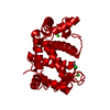
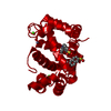
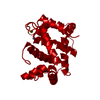
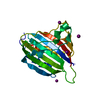
| Entry | Database: PDB / ID: 3psr | ||||||
|---|---|---|---|---|---|---|---|
| Title | HUMAN PSORIASIN (S100A7) CA2+ BOUND FORM (CRYSTAL FORM I) | ||||||
 Components Components | PSORIASIN | ||||||
 Keywords Keywords |  EF-HAND PROTEIN / CA-BINDING / PSORIASIS / S100 PROTEIN FAMILY EF-HAND PROTEIN / CA-BINDING / PSORIASIS / S100 PROTEIN FAMILY | ||||||
| Function / homology |  Function and homology information Function and homology informationpositive regulation of granulocyte chemotaxis / zinc ion sequestering activity / Metal sequestration by antimicrobial proteins / positive regulation of T cell chemotaxis / RAGE receptor binding / positive regulation of monocyte chemotaxis / epidermis development / keratinocyte differentiation / response to reactive oxygen species / calcium-dependent protein binding ...positive regulation of granulocyte chemotaxis / zinc ion sequestering activity / Metal sequestration by antimicrobial proteins / positive regulation of T cell chemotaxis / RAGE receptor binding / positive regulation of monocyte chemotaxis / epidermis development / keratinocyte differentiation / response to reactive oxygen species / calcium-dependent protein binding / azurophil granule lumen / antimicrobial humoral immune response mediated by antimicrobial peptide / angiogenesis / collagen-containing extracellular matrix / response to lipopolysaccharide / positive regulation of ERK1 and ERK2 cascade / focal adhesion / calcium ion binding / Neutrophil degranulation / endoplasmic reticulum / extracellular space / zinc ion binding / extracellular region / nucleus / cytosol / cytoplasmSimilarity search - Function | ||||||
| Biological species |  Homo sapiens (human) Homo sapiens (human) | ||||||
| Method | X-RAY DIFFRACTION / MOLECULAR REPLACEMENT / Resolution: 2.5 Å | ||||||
 Authors Authors | Brodersen, D.E. / Nyborg, J. / Kjeldgaard, M. | ||||||
 Citation Citation | Journal: Biochemistry / Year: 1999 Title: Zinc-binding site of an S100 protein revealed. Two crystal structures of Ca2+-bound human psoriasin (S100A7) in the Zn2+-loaded and Zn2+-free states. Authors: Brodersen, D.E. / Nyborg, J. / Kjeldgaard, M. #1: Journal: Structure / Year: 1998Title: EF-Hands at Atomic Resolution: The Structure of Human Psoriasin (S100A7) Solved by MAD Phasing Authors: Brodersen, D.E. / Etzerodt, M. / Madsen, P. / Celis, J.E. / Thogersen, H.C. / Nyborg, J. / Kjeldgaard, M. #2: Journal: Acta Crystallogr.,Sect.D / Year: 1997Title: Crystallization and Preliminary X-Ray Diffraction Studies of Psoriasin Authors: Nolsoe, S. / Thirup, S. / Etzerodt, M. / Thogersen, H.C. / Nyborg, J. #3: Journal: J.Invest.Dermatol. / Year: 1991Title: Molecular Cloning, Occurrence, and Expression of a Novel Partially Secreted Protein "Psoriasin" that is Highly Up-Regulated in Psoriatic Skin Authors: Madsen, P. / Rasmussen, H.H. / Leffers, H. / Honore, B. / Dejgaard, K. / Olsen, E. / Kiil, J. / Walbum, E. / Andersen, A.H. / Basse, B. / Lauridsen, J.B. / Ratz, G.P. / Celis, A. / ...Authors: Madsen, P. / Rasmussen, H.H. / Leffers, H. / Honore, B. / Dejgaard, K. / Olsen, E. / Kiil, J. / Walbum, E. / Andersen, A.H. / Basse, B. / Lauridsen, J.B. / Ratz, G.P. / Celis, A. / Vandekerckhove, J. / Celis, J.E. | ||||||
| History |
|
- Structure visualization
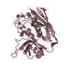
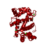
| Structure viewer | Molecule: MolmilJmol/JSmol |
|---|
- Downloads & links
-Download
| PDBx/mmCIF format | 3psr.cif.gz | 49.9 KB | Display | PDBx/mmCIF format |
|---|---|---|---|---|
| PDB format | pdb3psr.ent.gz | 38.3 KB | Display | PDB format |
| PDBx/mmJSON format | 3psr.json.gz | Tree view | PDBx/mmJSON format | |
| Others |  Other downloads Other downloads |
-Validation report
| Arichive directory | https://data.pdbj.org/pub/pdb/validation_reports/ps/3psrftp://data.pdbj.org/pub/pdb/validation_reports/ps/3psr | HTTPS FTP |
|---|
-Related structure data
| Related structure data | 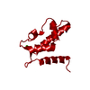 2psrC  1psrS S: Starting model for refinement C: citing same article ( |
|---|---|
| Similar structure data |


-Links
 PDBj
PDBj
- Assembly
| Deposited unit | 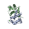
| ||||||||
|---|---|---|---|---|---|---|---|---|---|
| 1 |
| ||||||||
| Unit cell |
| ||||||||
| Noncrystallographic symmetry (NCS) | NCS oper: (Code: given Matrix: (0.1597, 0.9314, 0.3272), Vector : |
-Components
| #1: Protein | Mass: 11343.784 Da / Num. of mol.: 2 Source method: isolated from a genetically manipulated source Details: CHAIN A IS CA2+ AND ZN2+ BOUND FORM, CHAIN B IS CA2+ BOUND FORM Source: (gene. exp.) Homo sapiens (human) / Cell: KERATINOCYTESCellular location: CYTOPLASMIC, OR MAY BE SECRETED BY A NON-CLASSICAL SECRETORY PATHWAY CytoplasmPlasmid: PT7H6FX-PS.4 / Species (production host): Escherichia coli / Production host:  Escherichia coli BL21 (bacteria) / Strain (production host): BL21 / References: UniProt: P31151 Escherichia coli BL21 (bacteria) / Strain (production host): BL21 / References: UniProt: P31151#2: Chemical |   Mass: 40.078 Da / Num. of mol.: 2 / Source method: obtained synthetically / Formula: Ca Mass: 40.078 Da / Num. of mol.: 2 / Source method: obtained synthetically / Formula: Ca#3: Chemical | ChemComp-ZN / |   Mass: 65.409 Da / Num. of mol.: 1 / Source method: obtained synthetically / Formula: Zn Mass: 65.409 Da / Num. of mol.: 1 / Source method: obtained synthetically / Formula: Zn#4: Water | ChemComp-HOH / | Water Mass: 18.015 Da / Num. of mol.: 70 / Source method: isolated from a natural source / Formula: H2O Mass: 18.015 Da / Num. of mol.: 70 / Source method: isolated from a natural source / Formula: H2OCompound details | FOR THIS STRUCTURE, ONLY ONE OF THE TWO ZINC BINDING SITES ACTUALLY BINDS ZINC (MONOMER A), WHEREAS ...FOR THIS STRUCTURE, ONLY ONE OF THE TWO ZINC BINDING SITES ACTUALLY BINDS ZINC (MONOMER A), WHEREAS THE OTHER (MONOMER B) DOES NOT. ONLY ONE ZINC SITE IS LISTED ON SITE RECORDS BELOW. | |
|---|
-Experimental details
-Experiment
| Experiment | Method: X-RAY DIFFRACTION / Number of used crystals: 1 |
|---|
- Sample preparation
| Crystal | Density Matthews: 2.22 Å3/Da / Density % sol: 44.7 % | ||||||||||||||||||||||||||||||
|---|---|---|---|---|---|---|---|---|---|---|---|---|---|---|---|---|---|---|---|---|---|---|---|---|---|---|---|---|---|---|---|
| Crystal grow | pH: 7.7 / Details: pH 7.7 | ||||||||||||||||||||||||||||||
| Crystal grow | *PLUS Temperature: 20 ℃ / Method: unknown / PH range low: 8 / PH range high: 6.5 | ||||||||||||||||||||||||||||||
| Components of the solutions | *PLUS
|
-Data collection
| Diffraction | Mean temperature: 298 K |
|---|---|
| Diffraction source | Source: ROTATING ANODE / Type: RIGAKU RUH2R / Wavelength: 1.5418 |
| Detector | Type: RIGAKU RAXIS II / Detector: IMAGE PLATE |
| Radiation | Monochromatic (M) / Laue (L): M / Scattering type: x-ray |
| Radiation wavelength | Wavelength: 1.5418 Å / Relative weight: 1 |
| Reflection | Resolution: 2.5→34.3 Å / Num. obs: 8199 / % possible obs: 99.3 % / Redundancy: 5.4 % / Rsym value: 0.122 / Net I/σ(I): 5.4 |
| Reflection shell | Resolution: 2.5→2.64 Å / Redundancy: 5.3 % / Mean I/σ(I) obs: 1.4 / Rsym value: 0.544 / % possible all: 99.3 |
| Reflection | *PLUS Rmerge(I) obs: 0.122 |
| Reflection shell | *PLUS % possible obs: 99.3 % / Rmerge(I) obs: 0.544 |
- Processing
| Software |
| |||||||||||||||||||||||||||||||||
|---|---|---|---|---|---|---|---|---|---|---|---|---|---|---|---|---|---|---|---|---|---|---|---|---|---|---|---|---|---|---|---|---|---|---|
| Refinement | Method to determine structure: MOLECULAR REPLACEMENT Starting model: 1PSR Resolution: 2.5→100 Å / Num. parameters: 6450 / Num. restraintsaints: 8061 / Cross valid method: FREE R / Stereochemistry target values: ENGH AND HUBER
| |||||||||||||||||||||||||||||||||
| Solvent computation | Solvent model: MOEWS & KRETSINGER, J.MOL.BIOL. 91(1973) 201-228 | |||||||||||||||||||||||||||||||||
| Refine analyze | Num. disordered residues: 0 / Occupancy sum hydrogen: 0 / Occupancy sum non hydrogen: 1610.3 | |||||||||||||||||||||||||||||||||
| Refinement step | Cycle: LAST / Resolution: 2.5→100 Å
| |||||||||||||||||||||||||||||||||
| Refine LS restraints |
| |||||||||||||||||||||||||||||||||
| Software | *PLUS Name: SHELXL-97 / Classification: refinement | |||||||||||||||||||||||||||||||||
| Refinement | *PLUS Rfactor obs: 0.224 / Rfactor Rfree: 0.293 | |||||||||||||||||||||||||||||||||
| Solvent computation | *PLUS | |||||||||||||||||||||||||||||||||
| Displacement parameters | *PLUS |
