 Movie
Movie Controller

[English] 日本語
 Yorodumi
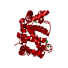
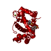
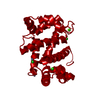
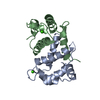
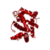
- PDB-2psr: HUMAN PSORIASIN (S100A7) CA2+ AND ZN2+ BOUND FORM (CRYSTAL FORM II) -
+ Open data

- Basic information
| Entry | Database: PDB / ID: 2psr | ||||||
|---|---|---|---|---|---|---|---|
| Title | HUMAN PSORIASIN (S100A7) CA2+ AND ZN2+ BOUND FORM (CRYSTAL FORM II) | ||||||
 Components Components | PSORIASIN | ||||||
 Keywords Keywords |  EF-HAND PROTEIN / CA-BINDING / ZN-BINDING / PSORIASIS / S100 PROTEIN FAMILY EF-HAND PROTEIN / CA-BINDING / ZN-BINDING / PSORIASIS / S100 PROTEIN FAMILY | ||||||
| Function / homology |  Function and homology information Function and homology informationpositive regulation of granulocyte chemotaxis / zinc ion sequestering activity / Metal sequestration by antimicrobial proteins / positive regulation of T cell chemotaxis / RAGE receptor binding / positive regulation of monocyte chemotaxis / epidermis development / keratinocyte differentiation / response to reactive oxygen species / calcium-dependent protein binding ...positive regulation of granulocyte chemotaxis / zinc ion sequestering activity / Metal sequestration by antimicrobial proteins / positive regulation of T cell chemotaxis / RAGE receptor binding / positive regulation of monocyte chemotaxis / epidermis development / keratinocyte differentiation / response to reactive oxygen species / calcium-dependent protein binding / azurophil granule lumen / antimicrobial humoral immune response mediated by antimicrobial peptide / angiogenesis / collagen-containing extracellular matrix / response to lipopolysaccharide / positive regulation of ERK1 and ERK2 cascade / focal adhesion / calcium ion binding / Neutrophil degranulation / endoplasmic reticulum / extracellular space / zinc ion binding / extracellular region / nucleus / cytosol / cytoplasmSimilarity search - Function | ||||||
| Biological species |  Homo sapiens (human) Homo sapiens (human) | ||||||
| Method | X-RAY DIFFRACTION / SYNCHROTRON / MOLECULAR REPLACEMENT / Resolution: 2.05 Å | ||||||
 Authors Authors | Brodersen, D.E. / Nyborg, J. / Kjeldgaard, M. | ||||||
 Citation Citation | Journal: Biochemistry / Year: 1999 Title: Zinc-binding site of an S100 protein revealed. Two crystal structures of Ca2+-bound human psoriasin (S100A7) in the Zn2+-loaded and Zn2+-free states. Authors: Brodersen, D.E. / Nyborg, J. / Kjeldgaard, M. #1: Journal: Structure / Year: 1998Title: EF-Hands at Atomic Resolution: The Structure of Human Psoriasin (S100A7) Solved by MAD Phasing Authors: Brodersen, D.E. / Etzerodt, M. / Madsen, P. / Celis, J.E. / Thogersen, H.C. / Nyborg, J. / Kjeldgaard, M. #2: Journal: Acta Crystallogr.,Sect.D / Year: 1997Title: Crystallization and Preliminary X-Ray Diffraction Studies of Psoriasin Authors: Nolsoe, S. / Thirup, S. / Etzerodt, M. / Thogersen, H.C. / Nyborg, J. #3: Journal: J.Invest.Dermatol. / Year: 1991Title: Molecular Cloning, Occurrence, and Expression of a Novel Partially Secreted Protein "Psoriasin" that is Highly Up-Regulated in Psoriatic Skin Authors: Madsen, P. / Rasmussen, H.H. / Leffers, H. / Honore, B. / Dejgaard, K. / Olsen, E. / Kiil, J. / Walbum, E. / Andersen, A.H. / Basse, B. / Lauridsen, J.B. / Ratz, G.P. / Celis, A. / ...Authors: Madsen, P. / Rasmussen, H.H. / Leffers, H. / Honore, B. / Dejgaard, K. / Olsen, E. / Kiil, J. / Walbum, E. / Andersen, A.H. / Basse, B. / Lauridsen, J.B. / Ratz, G.P. / Celis, A. / Vandekerckhove, J. / Celis, J.E. | ||||||
| History |
|
- Structure visualization
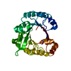
| Structure viewer | Molecule: MolmilJmol/JSmol |
|---|
- Downloads & links
-Download
| PDBx/mmCIF format | 2psr.cif.gz | 31.6 KB | Display | PDBx/mmCIF format |
|---|---|---|---|---|
| PDB format | pdb2psr.ent.gz | 23 KB | Display | PDB format |
| PDBx/mmJSON format | 2psr.json.gz | Tree view | PDBx/mmJSON format | |
| Others |  Other downloads Other downloads |
-Validation report
| Arichive directory | https://data.pdbj.org/pub/pdb/validation_reports/ps/2psrftp://data.pdbj.org/pub/pdb/validation_reports/ps/2psr | HTTPS FTP |
|---|
-Related structure data
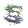
| Related structure data |  3psrC  1psrS S: Starting model for refinement C: citing same article ( |
|---|---|
| Similar structure data |


-Links
 PDBj
PDBj
- Assembly
| Deposited unit | 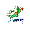
| ||||||||
|---|---|---|---|---|---|---|---|---|---|
| 1 | 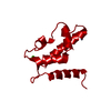
| ||||||||
| Unit cell |
|
-Components
| #1: Protein | Mass: 11343.784 Da / Num. of mol.: 1 Source method: isolated from a genetically manipulated source Details: CA2+ AND ZN2+ BOUND FORM / Source: (gene. exp.) Homo sapiens (human) / Cell: KERATINOCYTESCellular location: CYTOPLASMIC, OR MAY BE SECRETED BY A NON-CLASSICAL SECRETORY PATHWAY CytoplasmPlasmid: PT7H6FX-PS.4 / Species (production host): Escherichia coli / Production host:  Escherichia coli BL21 (bacteria) / Strain (production host): BL21 / References: UniProt: P31151 Escherichia coli BL21 (bacteria) / Strain (production host): BL21 / References: UniProt: P31151 |
|---|---|
| #2: Chemical | ChemComp-CA /   Mass: 40.078 Da / Num. of mol.: 1 / Source method: obtained synthetically / Formula: Ca Mass: 40.078 Da / Num. of mol.: 1 / Source method: obtained synthetically / Formula: Ca |
| #3: Chemical | ChemComp-ZN /   Mass: 65.409 Da / Num. of mol.: 1 / Source method: obtained synthetically / Formula: Zn Mass: 65.409 Da / Num. of mol.: 1 / Source method: obtained synthetically / Formula: Zn |
| #4: Water | ChemComp-HOH / Water Mass: 18.015 Da / Num. of mol.: 106 / Source method: isolated from a natural source / Formula: H2O Mass: 18.015 Da / Num. of mol.: 106 / Source method: isolated from a natural source / Formula: H2O |
-Experimental details
-Experiment
| Experiment | Method: X-RAY DIFFRACTION / Number of used crystals: 1 |
|---|
- Sample preparation
| Crystal | Density Matthews: 3.06 Å3/Da / Density % sol: 59.7 % | ||||||||||||||||||||||||||||||
|---|---|---|---|---|---|---|---|---|---|---|---|---|---|---|---|---|---|---|---|---|---|---|---|---|---|---|---|---|---|---|---|
| Crystal grow | pH: 6.7 / Details: pH 6.7 | ||||||||||||||||||||||||||||||
| Crystal grow | *PLUS Temperature: 4 ℃ / Method: unknown / Details: or 20 degrees centigrade | ||||||||||||||||||||||||||||||
| Components of the solutions | *PLUS
|
-Data collection
| Diffraction | Mean temperature: 100 K |
|---|---|
| Diffraction source | Source: SYNCHROTRON / Site: EMBL/DESY, HAMBURG  / Beamline: BW7B / Wavelength: 0.862 / Beamline: BW7B / Wavelength: 0.862 |
| Detector | Type: MARRESEARCH / Detector: IMAGE PLATE / Date: Sep 1, 1995 |
| Radiation | Monochromatic (M) / Laue (L): M / Scattering type: x-ray |
| Radiation wavelength | Wavelength: 0.862 Å / Relative weight: 1 |
| Reflection | Resolution: 2.05→20.1 Å / Num. obs: 10791 / % possible obs: 98.9 % / Observed criterion σ(I): 0 / Redundancy: 7 % / Rsym value: 0.113 / Net I/σ(I): 15.9 |
| Reflection shell | Resolution: 2.05→2.12 Å / Redundancy: 6.9 % / Mean I/σ(I) obs: 5 / Rsym value: 0.423 / % possible all: 100 |
| Reflection | *PLUS Rmerge(I) obs: 0.113 |
| Reflection shell | *PLUS % possible obs: 100 % / Rmerge(I) obs: 0.423 |
- Processing
| Software |
| |||||||||||||||||||||||||||||||||
|---|---|---|---|---|---|---|---|---|---|---|---|---|---|---|---|---|---|---|---|---|---|---|---|---|---|---|---|---|---|---|---|---|---|---|
| Refinement | Method to determine structure: MOLECULAR REPLACEMENT Starting model: 1PSR Resolution: 2.05→100 Å / Num. parameters: 3513 / Num. restraintsaints: 3122 / Cross valid method: FREE R / Stereochemistry target values: ENGH AND HUBER
| |||||||||||||||||||||||||||||||||
| Solvent computation | Solvent model: MOEWS & KRETSINGER, J.MOL.BIOL. 91(1973) 201-228 | |||||||||||||||||||||||||||||||||
| Refine analyze | Num. disordered residues: 0 / Occupancy sum hydrogen: 0 / Occupancy sum non hydrogen: 876.89 | |||||||||||||||||||||||||||||||||
| Refinement step | Cycle: LAST / Resolution: 2.05→100 Å
| |||||||||||||||||||||||||||||||||
| Refine LS restraints |
| |||||||||||||||||||||||||||||||||
| Software | *PLUS Name: SHELXL-97 / Classification: refinement | |||||||||||||||||||||||||||||||||
| Refinement | *PLUS Num. reflection all: 10236 / Rfactor all: 0.219 / Rfactor obs: 0.27 | |||||||||||||||||||||||||||||||||
| Solvent computation | *PLUS | |||||||||||||||||||||||||||||||||
| Displacement parameters | *PLUS |
