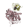
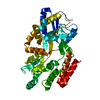
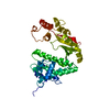
1.6 Angstrom resolution crystal structure of putative streptothricin acetyltransferase from Bacillus anthracis str. Ames in complex with acetyl coenzyme A
Components
Putative streptothricin acetyltransferase
Keywords
TRANSFERASE / Putative Streptothricin Acetyltransferase / Toxin Production and Resistance / Infectious Diseases / Structural Genomics / Center for Structural Genomics of Infectious Diseases (CSGID)
Resolution: 1.6→30 Å / Num. all: 70883 / Num. obs: 70883 / % possible obs: 98.5 % / Observed criterion σ(I): -3 / Redundancy: 5.4 % / Biso Wilson estimate: 21 Å2 / Rmerge(I) obs: 0.065 / Net I/σ(I): 20.6
Reflection shell
Resolution: 1.6→1.63 Å / Redundancy: 4.5 % / Rmerge(I) obs: 0.489 / Mean I/σ(I) obs: 2.96 / Num. unique all: 2912 / % possible all: 82
-
Processing
Software
Name
Version
Classification
Blu-Ice
Max
datacollection
PHENIX
modelbuilding
REFMAC
5.5.0102
refinement
HKL-2000
datareduction
HKL-2000
datascaling
PHENIX
phasing
Refinement
Method to determine structure: SAD Starting model: SAD model of the data set collected on 21-ID-F Resolution: 1.6→29.28 Å / Cor.coef. Fo:Fc: 0.96 / Cor.coef. Fo:Fc free: 0.945 / SU B: 3.794 / SU ML: 0.068 / Isotropic thermal model: isotropic / Cross valid method: THROUGHOUT / ESU R Free: 0.101 / Stereochemistry target values: MAXIMUM LIKELIHOOD / Details: HYDROGENS HAVE BEEN ADDED IN THE RIDING POSITIONS
Rfactor
Num. reflection
% reflection
Selection details
Rfree
0.22203
3571
5 %
RANDOM
Rwork
0.18863
-
-
-
obs
0.1903
67311
98.44 %
-
all
-
67311
-
-
Solvent computation
Ion probe radii: 0.8 Å / Shrinkage radii: 0.8 Å / VDW probe radii: 1.2 Å / Solvent model: BABINET MODEL WITH MASK
Displacement parameters
Biso mean: 28.639 Å2
Baniso -1
Baniso -2
Baniso -3
1-
0.2 Å2
0 Å2
-1.19 Å2
2-
-
-0.19 Å2
0 Å2
3-
-
-
-0.33 Å2
Refinement step
Cycle: LAST / Resolution: 1.6→29.28 Å
Protein
Nucleic acid
Ligand
Solvent
Total
Num. atoms
4308
0
180
355
4843
Refine LS restraints
Refine-ID
Type
Dev ideal
Dev ideal target
Number
X-RAY DIFFRACTION
r_bond_refined_d
0.01
0.022
5013
X-RAY DIFFRACTION
r_bond_other_d
0.001
0.02
3293
X-RAY DIFFRACTION
r_angle_refined_deg
1.591
1.992
6854
X-RAY DIFFRACTION
r_angle_other_deg
0.904
3
8080
X-RAY DIFFRACTION
r_dihedral_angle_1_deg
2.306
5
597
X-RAY DIFFRACTION
r_dihedral_angle_2_deg
21.727
25.301
249
X-RAY DIFFRACTION
r_dihedral_angle_3_deg
8.071
15
874
X-RAY DIFFRACTION
r_dihedral_angle_4_deg
11.293
15
17
X-RAY DIFFRACTION
r_chiral_restr
0.103
0.2
719
X-RAY DIFFRACTION
r_gen_planes_refined
0.006
0.02
5606
X-RAY DIFFRACTION
r_gen_planes_other
0.002
0.02
1023
X-RAY DIFFRACTION
r_mcbond_it
0.796
1.5
2862
X-RAY DIFFRACTION
r_mcbond_other
0.202
1.5
1146
X-RAY DIFFRACTION
r_mcangle_it
1.469
2
4673
X-RAY DIFFRACTION
r_scbond_it
2.366
3
2151
X-RAY DIFFRACTION
r_scangle_it
3.69
4.5
2181
LS refinement shell
Resolution: 1.599→1.641 Å / Total num. of bins used: 20
Rfactor
Num. reflection
% reflection
Rfree
0.251
218
-
Rwork
0.229
4139
-
obs
-
4139
82.88 %
Refinement TLS params.
Method: refined / Refine-ID: X-RAY DIFFRACTION
ID
L11 (°2)
L12 (°2)
L13 (°2)
L22 (°2)
L23 (°2)
L33 (°2)
S11 (Å °)
S12 (Å °)
S13 (Å °)
S21 (Å °)
S22 (Å °)
S23 (Å °)
S31 (Å °)
S32 (Å °)
S33 (Å °)
T11 (Å2)
T12 (Å2)
T13 (Å2)
T22 (Å2)
T23 (Å2)
T33 (Å2)
Origin x (Å)
Origin y (Å)
Origin z (Å)
1
0.8233
-0.4361
0.102
1.3451
0.4933
0.6087
0.0465
-0.0039
-0.0321
0.0052
-0.0007
-0.0389
-0.0224
0.0427
-0.0459
0.0779
0.0104
0.0451
0.111
0.024
0.1158
35.2751
25.2927
11.1887
2
1.4107
-0.4072
0.3298
1.8606
0.2651
1.1962
0.005
-0.0776
0.0393
0.1301
0.0018
0.122
-0.0086
-0.0311
-0.0068
0.0213
0.0052
0.0317
0.0352
0.0026
0.0549
29.7312
29.7917
16.3385
3
2.2815
0.1063
-0.5722
1.869
-0.1588
3.2509
0.0371
0.0762
-0.0195
0.0674
-0.0383
-0.0594
0.1846
0.1115
0.0012
0.0169
0.0106
0.0093
0.044
0.0099
0.1307
28.9802
18.7619
4.7137
4
0.6447
-0.2846
-0.4145
1.7745
1.28
2.7113
0.0515
-0.0203
0.0106
-0.1446
0.1062
-0.2062
-0.1896
0.0325
-0.1577
0.0918
0.0274
-0.0023
0.1383
-0.0083
0.158
4.9353
23.9518
38.6786
5
0.9404
0.025
0.8019
1.1843
0.3252
3.697
0.0878
-0.0907
-0.0129
0.129
-0.0026
-0.0811
0.2658
0.0054
-0.0853
0.1047
0.0138
-0.0142
0.0799
-0.0235
0.0684
4.0614
19.6637
42.1504
6
1.5361
-0.6194
0.7536
0.761
0.1188
4.3581
0.0313
-0.0522
0.0917
-0.0577
-0.1393
0.0133
-0.148
-0.323
0.108
0.0817
0.013
0.0111
0.1481
-0.0236
0.0288
1.8376
27.1909
52.8796
7
2.735
0.2124
-0.5118
1.8838
-0.1634
1.8393
0.0119
0.1629
-0.324
0.1415
0.1309
-0.401
0.22
0.0976
-0.1428
0.2529
0.0156
-0.0668
0.1149
-0.1044
0.2227
17.04
14.3922
72.2021
8
1.925
0.1693
-0.0116
1.7867
0.4189
1.8633
0.0544
0.1375
-0.0847
0.1577
0.1798
-0.3595
0.1737
0.2609
-0.2343
0.0979
0.0298
-0.0368
0.0952
-0.0715
0.1076
21.2412
20.3296
72.4885
9
1.8868
0.5992
0.8049
2.746
-0.2926
3.777
-0.0054
0.0566
-0.1189
-0.0496
0.1097
-0.042
0.321
-0.2111
-0.1043
0.0753
-0.0154
0.0167
0.0726
-0.0183
0.0285
7.449
22.5093
67.6287
Refinement TLS group
ID
Refine-ID
Refine TLS-ID
Auth asym-ID
Auth seq-ID
1
X-RAY DIFFRACTION
1
A
2 - 67
2
X-RAY DIFFRACTION
2
A
68 - 148
3
X-RAY DIFFRACTION
3
A
149 - 182
4
X-RAY DIFFRACTION
4
B
-1 - 36
5
X-RAY DIFFRACTION
5
B
37 - 137
6
X-RAY DIFFRACTION
6
B
138 - 183
7
X-RAY DIFFRACTION
7
C
1 - 72
8
X-RAY DIFFRACTION
8
C
73 - 148
9
X-RAY DIFFRACTION
9
C
149 - 183
+
About Yorodumi
-
News
-
Feb 9, 2022. New format data for meta-information of EMDB entries
New format data for meta-information of EMDB entries
Version 3 of the EMDB header file is now the official format.
The previous official version 1.9 will be removed from the archive.
In the structure databanks used in Yorodumi, some data are registered as the other names, "COVID-19 virus" and "2019-nCoV". Here are the details of the virus and the list of structure data.
Jan 31, 2019. EMDB accession codes are about to change! (news from PDBe EMDB page)
EMDB accession codes are about to change! (news from PDBe EMDB page)
The allocation of 4 digits for EMDB accession codes will soon come to an end. Whilst these codes will remain in use, new EMDB accession codes will include an additional digit and will expand incrementally as the available range of codes is exhausted. The current 4-digit format prefixed with “EMD-” (i.e. EMD-XXXX) will advance to a 5-digit format (i.e. EMD-XXXXX), and so on. It is currently estimated that the 4-digit codes will be depleted around Spring 2019, at which point the 5-digit format will come into force.
The EM Navigator/Yorodumi systems omit the EMD- prefix.
Related info.:Q: What is EMD? / ID/Accession-code notation in Yorodumi/EM Navigator
Yorodumi is a browser for structure data from EMDB, PDB, SASBDB, etc.
This page is also the successor to EM Navigator detail page, and also detail information page/front-end page for Omokage search.
The word "yorodu" (or yorozu) is an old Japanese word meaning "ten thousand". "mi" (miru) is to see.
Related info.:EMDB / PDB / SASBDB / Comparison of 3 databanks / Yorodumi Search / Aug 31, 2016. New EM Navigator & Yorodumi / Yorodumi Papers / Jmol/JSmol / Function and homology information / Changes in new EM Navigator and Yorodumi
 Movie
Movie Controller

 Yorodumi
 Open data
 Basic information
 Components
 Keywords
 TRANSFERASE / Putative Streptothricin Acetyltransferase / Toxin Production and Resistance /
  Function and homology information
 Authors
 Citation
 Structure visualization
 Downloads & links
 Other downloads


 PDBj
PDBj

 Assembly


 Mass: 809.571 Da / Num. of mol.: 3 / Source method: obtained synthetically / Formula: C23H38N7O17P3S
 Mass: 94.971 Da / Num. of mol.: 5 / Source method: obtained synthetically / Formula: PO4
 Mass: 39.098 Da / Num. of mol.: 2 / Source method: obtained synthetically / Formula: K
 Mass: 18.015 Da / Num. of mol.: 355 / Source method: isolated from a natural source / Formula: H2O
 Sample preparation
 Processing