 Movie
Movie Controller

[English] 日本語
 Yorodumi
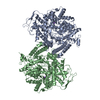
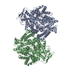
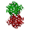
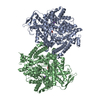
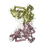
- PDB-3pfl: CRYSTAL STRUCTURE OF PFL FROM E.COLI IN COMPLEX WITH SUBSTRATE AN... -
+ Open data

- Basic information
| Entry | Database: PDB / ID: 3pfl | ||||||
|---|---|---|---|---|---|---|---|
| Title | CRYSTAL STRUCTURE OF PFL FROM E.COLI IN COMPLEX WITH SUBSTRATE ANALOGUE OXAMATE | ||||||
 Components Components | PROTEIN (FORMATE ACETYLTRANSFERASE 1) | ||||||
 Keywords Keywords | LYASE/TRANSFERASE / GLYCYL RADICAL ENZYME /  TRANSFERASE / GLUCOSE METABOLISM / LYASE-TRANSFERASE COMPLEX TRANSFERASE / GLUCOSE METABOLISM / LYASE-TRANSFERASE COMPLEX | ||||||
| Function / homology |  Function and homology informationformate C-acetyltransferase / formate C-acetyltransferase activity / glucose metabolic process / membrane / cytosol Function and homology informationformate C-acetyltransferase / formate C-acetyltransferase activity / glucose metabolic process / membrane / cytosolSimilarity search - Function | ||||||
| Biological species |  Escherichia coli (E. coli) Escherichia coli (E. coli) | ||||||
| Method | X-RAY DIFFRACTION / SYNCHROTRON / MOLECULAR REPLACEMENT / Resolution: 2.6 Å | ||||||
 Authors Authors | Becker, A. / Fritz-Wolf, K. / Kabsch, W. / Knappe, J. / Schultz, S. / Wagner, A.F.V. | ||||||
 Citation Citation | Journal: Nat.Struct.Biol. / Year: 1999 Title: Structure and mechanism of the glycyl radical enzyme pyruvate formate-lyase. Authors: Becker, A. / Fritz-Wolf, K. / Kabsch, W. / Knappe, J. / Schultz, S. / Volker Wagner, A.F. | ||||||
| History |
|
- Structure visualization
| Structure viewer | Molecule: MolmilJmol/JSmol |
|---|
- Downloads & links
-Download
| PDBx/mmCIF format | 3pfl.cif.gz | 308.2 KB | Display | PDBx/mmCIF format |
|---|---|---|---|---|
| PDB format | pdb3pfl.ent.gz | 250.8 KB | Display | PDB format |
| PDBx/mmJSON format | 3pfl.json.gz | Tree view | PDBx/mmJSON format | |
| Others |  Other downloads Other downloads |
-Validation report
| Arichive directory | https://data.pdbj.org/pub/pdb/validation_reports/pf/3pflftp://data.pdbj.org/pub/pdb/validation_reports/pf/3pfl | HTTPS FTP |
|---|
-Related structure data


-Links
 PDBj
PDBj
- Assembly
| Deposited unit | 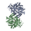
| ||||||||
|---|---|---|---|---|---|---|---|---|---|
| 1 |
| ||||||||
| Unit cell |
| ||||||||
| Noncrystallographic symmetry (NCS) | NCS oper: (Code: given Matrix: (-0.9119, 0.4058, 0.061), Vector : |
-Components
| #1: Protein | Mass: 85327.898 Da / Num. of mol.: 2 / Source method: isolated from a natural source / Source: (natural) Escherichia coli (E. coli) / References: UniProt: P09373, formate C-acetyltransferase#2: Chemical | Oxamic acid  Mass: 89.050 Da / Num. of mol.: 2 / Source method: obtained synthetically / Formula: C2H3NO3 Mass: 89.050 Da / Num. of mol.: 2 / Source method: obtained synthetically / Formula: C2H3NO3#3: Water | ChemComp-HOH / | Water Mass: 18.015 Da / Num. of mol.: 527 / Source method: isolated from a natural source / Formula: H2O Mass: 18.015 Da / Num. of mol.: 527 / Source method: isolated from a natural source / Formula: H2O |
|---|
-Experimental details
-Experiment
| Experiment | Method: X-RAY DIFFRACTION / Number of used crystals: 1 |
|---|
- Sample preparation
| Crystal | Density Matthews: 2.97 Å3/Da / Density % sol: 55 % | ||||||||||||||||||||||||||||||||||||||||||||||||||||||||||||||||||||||||||||||||||||
|---|---|---|---|---|---|---|---|---|---|---|---|---|---|---|---|---|---|---|---|---|---|---|---|---|---|---|---|---|---|---|---|---|---|---|---|---|---|---|---|---|---|---|---|---|---|---|---|---|---|---|---|---|---|---|---|---|---|---|---|---|---|---|---|---|---|---|---|---|---|---|---|---|---|---|---|---|---|---|---|---|---|---|---|---|---|
| Crystal grow | pH: 7.3 / Details: pH 7.3 | ||||||||||||||||||||||||||||||||||||||||||||||||||||||||||||||||||||||||||||||||||||
| Crystal grow | *PLUS Method: microdialysis | ||||||||||||||||||||||||||||||||||||||||||||||||||||||||||||||||||||||||||||||||||||
| Components of the solutions | *PLUS
|
-Data collection
| Diffraction | Mean temperature: 100 K |
|---|---|
| Diffraction source | Source: SYNCHROTRON / Site: EMBL/DESY, HAMBURG  / Beamline: BW7B / Wavelength: 0.8467 / Beamline: BW7B / Wavelength: 0.8467 |
| Detector | Type: MARRESEARCH / Date: Feb 1, 1999 |
| Radiation | Protocol: SINGLE WAVELENGTH / Monochromatic (M) / Laue (L): M / Scattering type: x-ray |
| Radiation wavelength | Wavelength: 0.8467 Å / Relative weight: 1 |
| Reflection | Resolution: 2.6→20 Å / Num. obs: 61282 / % possible obs: 96.1 % / Observed criterion σ(I): 0 / Redundancy: 5.4 % / Biso Wilson estimate: 56.8 Å2 / Rmerge(I) obs: 0.088 |
| Reflection | *PLUS Num. measured all: 333383 |
- Processing
| Software |
| ||||||||||||||||||||||||||||||||||||||||||||||||||||||||||||||||||||||||||||||||
|---|---|---|---|---|---|---|---|---|---|---|---|---|---|---|---|---|---|---|---|---|---|---|---|---|---|---|---|---|---|---|---|---|---|---|---|---|---|---|---|---|---|---|---|---|---|---|---|---|---|---|---|---|---|---|---|---|---|---|---|---|---|---|---|---|---|---|---|---|---|---|---|---|---|---|---|---|---|---|---|---|---|
| Refinement | Method to determine structure: MOLECULAR REPLACEMENT / Resolution: 2.6→50 Å / Rfactor Rfree error: 0.005 / Data cutoff high rms absF: 626424.91 / Isotropic thermal model: RESTRAINED / Cross valid method: THROUGHOUT / σ(F): 0 Details: DISORDERED REGIONS (RESIDUES A1-A3 AND B1-B3) WERE MODELED STEREOCHEMICALLY
| ||||||||||||||||||||||||||||||||||||||||||||||||||||||||||||||||||||||||||||||||
| Solvent computation | Solvent model: FLAT MODEL / Bsol: 37.33 Å2 / ksol: 0.314 e/Å3 | ||||||||||||||||||||||||||||||||||||||||||||||||||||||||||||||||||||||||||||||||
| Displacement parameters | Biso mean: 48.7 Å2
| ||||||||||||||||||||||||||||||||||||||||||||||||||||||||||||||||||||||||||||||||
| Refine analyze |
| ||||||||||||||||||||||||||||||||||||||||||||||||||||||||||||||||||||||||||||||||
| Refinement step | Cycle: LAST / Resolution: 2.6→50 Å
| ||||||||||||||||||||||||||||||||||||||||||||||||||||||||||||||||||||||||||||||||
| Refine LS restraints |
| ||||||||||||||||||||||||||||||||||||||||||||||||||||||||||||||||||||||||||||||||
| LS refinement shell | Resolution: 2.6→2.76 Å / Rfactor Rfree error: 0.016 / Total num. of bins used: 6
| ||||||||||||||||||||||||||||||||||||||||||||||||||||||||||||||||||||||||||||||||
| Xplor file |
| ||||||||||||||||||||||||||||||||||||||||||||||||||||||||||||||||||||||||||||||||
| Software | *PLUS Name: CNS / Version: 0.5 / Classification: refinement | ||||||||||||||||||||||||||||||||||||||||||||||||||||||||||||||||||||||||||||||||
| Refine LS restraints | *PLUS
|
