 Movie
Movie Controller

[English] 日本語
 Yorodumi
- PDB-3pfb: Crystal structure of the Lactobacillus johnsonii cinnamoyl estera... -
+ Open data

- Basic information
| Entry | Database: PDB / ID: 3pfb | ||||||
|---|---|---|---|---|---|---|---|
| Title | Crystal structure of the Lactobacillus johnsonii cinnamoyl esterase LJ0536 S106A mutant in complex with ethylferulate | ||||||
 Components Components | Cinnamoyl esterase | ||||||
 Keywords Keywords |  HYDROLASE / alpha/beta hydrolase fold / esterase / cinnamoyl/feruloyl esterase / hydroxycinammates / extracellular HYDROLASE / alpha/beta hydrolase fold / esterase / cinnamoyl/feruloyl esterase / hydroxycinammates / extracellular | ||||||
| Function / homology |  Function and homology informationHydrolases; Acting on ester bonds; Carboxylic-ester hydrolases / aminopeptidase activity Function and homology informationHydrolases; Acting on ester bonds; Carboxylic-ester hydrolases / aminopeptidase activitySimilarity search - Function | ||||||
| Biological species |  Lactobacillus johnsonii (bacteria) Lactobacillus johnsonii (bacteria) | ||||||
| Method | X-RAY DIFFRACTION / MOLECULAR REPLACEMENT / Resolution: 1.58 Å | ||||||
 Authors Authors | Stogios, P.J. / Lai, K.K. / Vu, C. / Xu, X. / Cui, H. / Molloy, S. / Gonzalez, C.F. / Yakunin, A. / Savchenko, A. | ||||||
 Citation Citation | Journal: Plos One / Year: 2011 Title: An Inserted alpha/beta Subdomain Shapes the Catalytic Pocket of Lactobacillus johnsonii Cinnamoyl Esterase Authors: Lai, K.K. / Stogios, P.J. / Vu, C. / Xu, X. / Cui, H. / Molloy, S. / Savchenko, A. / Yakunin, A. / Gonzalez, C.F. | ||||||
| History |
|
- Structure visualization
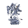
| Structure viewer | Molecule: MolmilJmol/JSmol |
|---|
- Downloads & links
-Download
| PDBx/mmCIF format | 3pfb.cif.gz | 247.9 KB | Display | PDBx/mmCIF format |
|---|---|---|---|---|
| PDB format | pdb3pfb.ent.gz | 199.4 KB | Display | PDB format |
| PDBx/mmJSON format | 3pfb.json.gz | Tree view | PDBx/mmJSON format | |
| Others |  Other downloads Other downloads |
-Validation report
| Arichive directory | https://data.pdbj.org/pub/pdb/validation_reports/pf/3pfbftp://data.pdbj.org/pub/pdb/validation_reports/pf/3pfb | HTTPS FTP |
|---|
-Related structure data
| Related structure data | 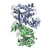 3pf8C 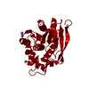 3pf9C 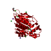 3pfcC 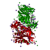 3qm1C  3s2zC  3pfa C: citing same article ( |
|---|---|
| Similar structure data |


-Links
 PDBj
PDBj
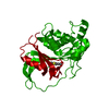
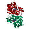
- Assembly
| Deposited unit | 
| ||||||||
|---|---|---|---|---|---|---|---|---|---|
| 1 |
| ||||||||
| Unit cell |
|
-Components
| #1: Protein | Mass: 30010.502 Da / Num. of mol.: 2 / Mutation: S106A Source method: isolated from a genetically manipulated source Source: (gene. exp.) Lactobacillus johnsonii (bacteria) / Gene: LJ0536 / Plasmid: p15TV-L / Production host: Escherichia coli (E. coli) / Strain (production host): BL21(DE3)References: UniProt: D3YEX6, Hydrolases; Acting on ester bonds; Carboxylic-ester hydrolases#2: Chemical | ChemComp-NH4 / Ammonium  Mass: 18.038 Da / Num. of mol.: 16 / Source method: obtained synthetically / Formula: H4N Mass: 18.038 Da / Num. of mol.: 16 / Source method: obtained synthetically / Formula: H4N#3: Chemical | ChemComp-CL / Chloride  Mass: 35.453 Da / Num. of mol.: 4 / Source method: obtained synthetically / Formula: Cl Mass: 35.453 Da / Num. of mol.: 4 / Source method: obtained synthetically / Formula: Cl#4: Chemical | 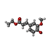  Mass: 222.237 Da / Num. of mol.: 2 / Source method: obtained synthetically / Formula: C12H14O4 Mass: 222.237 Da / Num. of mol.: 2 / Source method: obtained synthetically / Formula: C12H14O4#5: Water | ChemComp-HOH / | Water Mass: 18.015 Da / Num. of mol.: 907 / Source method: isolated from a natural source / Formula: H2O Mass: 18.015 Da / Num. of mol.: 907 / Source method: isolated from a natural source / Formula: H2O |
|---|
-Experimental details
-Experiment
| Experiment | Method: X-RAY DIFFRACTION / Number of used crystals: 1 |
|---|
- Sample preparation
| Crystal | Density Matthews: 2.22 Å3/Da / Density % sol: 44.66 % |
|---|---|
| Crystal grow | Temperature: 298 K / Method: vapor diffusion, sitting drop / pH: 8.5 Details: 0.1 M Tris pH 8.5, 0.2 M ammonium sulphate, 25% PEG3350, VAPOR DIFFUSION, SITTING DROP, temperature 298K |
-Data collection
| Diffraction | Mean temperature: 100 K | |||||||||||||||||||||||||||||||||||||||||||||||||||||||||||||||||||||||||||||||||||||||||||||||||||||||||||||||||||||||||||||||||||||||||||||||||||
|---|---|---|---|---|---|---|---|---|---|---|---|---|---|---|---|---|---|---|---|---|---|---|---|---|---|---|---|---|---|---|---|---|---|---|---|---|---|---|---|---|---|---|---|---|---|---|---|---|---|---|---|---|---|---|---|---|---|---|---|---|---|---|---|---|---|---|---|---|---|---|---|---|---|---|---|---|---|---|---|---|---|---|---|---|---|---|---|---|---|---|---|---|---|---|---|---|---|---|---|---|---|---|---|---|---|---|---|---|---|---|---|---|---|---|---|---|---|---|---|---|---|---|---|---|---|---|---|---|---|---|---|---|---|---|---|---|---|---|---|---|---|---|---|---|---|---|---|---|
| Diffraction source | Source: ROTATING ANODE / Type: RIGAKU FR-E SUPERBRIGHT / Wavelength: 1.5418 Å | |||||||||||||||||||||||||||||||||||||||||||||||||||||||||||||||||||||||||||||||||||||||||||||||||||||||||||||||||||||||||||||||||||||||||||||||||||
| Detector | Type: RIGAKU RAXIS HTC / Detector: IMAGE PLATE / Date: Dec 15, 2009 / Details: mirrors | |||||||||||||||||||||||||||||||||||||||||||||||||||||||||||||||||||||||||||||||||||||||||||||||||||||||||||||||||||||||||||||||||||||||||||||||||||
| Radiation | Monochromator: mirrors / Protocol: SINGLE WAVELENGTH / Monochromatic (M) / Laue (L): M / Scattering type: x-ray | |||||||||||||||||||||||||||||||||||||||||||||||||||||||||||||||||||||||||||||||||||||||||||||||||||||||||||||||||||||||||||||||||||||||||||||||||||
| Radiation wavelength | Wavelength: 1.5418 Å / Relative weight: 1 | |||||||||||||||||||||||||||||||||||||||||||||||||||||||||||||||||||||||||||||||||||||||||||||||||||||||||||||||||||||||||||||||||||||||||||||||||||
| Reflection | Resolution: 1.58→50 Å / Num. all: 52487 / Num. obs: 52487 / % possible obs: 73.1 % / Observed criterion σ(F): 0 / Observed criterion σ(I): -3 / Redundancy: 2 % / Rsym value: 0.046 / Net I/σ(I): 31.21 | |||||||||||||||||||||||||||||||||||||||||||||||||||||||||||||||||||||||||||||||||||||||||||||||||||||||||||||||||||||||||||||||||||||||||||||||||||
| Reflection shell |
|
- Processing
| Software |
| ||||||||||||||||||||||||||||||||||||||||||||||||||||||||||||||||||||||||||||||||||||||||||||||||||||||||||||||||||||||||||||||||||||||||||||||||||||||||||||||||||||||||||
|---|---|---|---|---|---|---|---|---|---|---|---|---|---|---|---|---|---|---|---|---|---|---|---|---|---|---|---|---|---|---|---|---|---|---|---|---|---|---|---|---|---|---|---|---|---|---|---|---|---|---|---|---|---|---|---|---|---|---|---|---|---|---|---|---|---|---|---|---|---|---|---|---|---|---|---|---|---|---|---|---|---|---|---|---|---|---|---|---|---|---|---|---|---|---|---|---|---|---|---|---|---|---|---|---|---|---|---|---|---|---|---|---|---|---|---|---|---|---|---|---|---|---|---|---|---|---|---|---|---|---|---|---|---|---|---|---|---|---|---|---|---|---|---|---|---|---|---|---|---|---|---|---|---|---|---|---|---|---|---|---|---|---|---|---|---|---|---|---|---|---|---|
| Refinement | Method to determine structure: MOLECULAR REPLACEMENT Starting model: Crystal structure of the Lactobacillus johnsonii cinnamoyl esterase LJ0536 S106A mutant Resolution: 1.58→44.2 Å / Cor.coef. Fo:Fc: 0.929 / Cor.coef. Fo:Fc free: 0.865 / SU B: 6.68 / SU ML: 0.109 / Cross valid method: THROUGHOUT / σ(F): 0 / ESU R Free: 0.17 / Stereochemistry target values: MAXIMUM LIKELIHOOD / Details: HYDROGENS HAVE BEEN ADDED IN THE RIDING POSITIONS
| ||||||||||||||||||||||||||||||||||||||||||||||||||||||||||||||||||||||||||||||||||||||||||||||||||||||||||||||||||||||||||||||||||||||||||||||||||||||||||||||||||||||||||
| Solvent computation | Ion probe radii: 0.8 Å / Shrinkage radii: 0.8 Å / VDW probe radii: 1.4 Å / Solvent model: BABINET MODEL WITH MASK | ||||||||||||||||||||||||||||||||||||||||||||||||||||||||||||||||||||||||||||||||||||||||||||||||||||||||||||||||||||||||||||||||||||||||||||||||||||||||||||||||||||||||||
| Displacement parameters | Biso mean: 26.599 Å2
| ||||||||||||||||||||||||||||||||||||||||||||||||||||||||||||||||||||||||||||||||||||||||||||||||||||||||||||||||||||||||||||||||||||||||||||||||||||||||||||||||||||||||||
| Refinement step | Cycle: LAST / Resolution: 1.58→44.2 Å
| ||||||||||||||||||||||||||||||||||||||||||||||||||||||||||||||||||||||||||||||||||||||||||||||||||||||||||||||||||||||||||||||||||||||||||||||||||||||||||||||||||||||||||
| Refine LS restraints |
| ||||||||||||||||||||||||||||||||||||||||||||||||||||||||||||||||||||||||||||||||||||||||||||||||||||||||||||||||||||||||||||||||||||||||||||||||||||||||||||||||||||||||||
| LS refinement shell | Resolution: 1.58→1.621 Å / Total num. of bins used: 20
| ||||||||||||||||||||||||||||||||||||||||||||||||||||||||||||||||||||||||||||||||||||||||||||||||||||||||||||||||||||||||||||||||||||||||||||||||||||||||||||||||||||||||||
| Refinement TLS params. | L11: 0 °2 / L12: 0 °2 / L13: 0 °2 / L22: 0 °2 / L23: 0 °2 / L33: 0 °2 / S11: 0 Å ° / S12: 0 Å ° / S13: 0 Å ° / S21: 0 Å ° / S22: 0 Å ° / S23: 0 Å ° / S31: 0 Å ° / S32: 0 Å ° / S33: -0 Å ° / T11: 0 Å2 / T12: 0 Å2 / T13: 0 Å2 / T22: 0 Å2 / T23: 0 Å2 / T33: 0 Å2 / Method: refined / Refine-ID: X-RAY DIFFRACTION
| ||||||||||||||||||||||||||||||||||||||||||||||||||||||||||||||||||||||||||||||||||||||||||||||||||||||||||||||||||||||||||||||||||||||||||||||||||||||||||||||||||||||||||
| Refinement TLS group |
|
